

Diagnosis of primary biliary melanoma with distinct imaging features: a case report and literature review
- PMID: 36999675
- PMCID: PMC10068995
- DOI: 10.1177/03000605231164005
Diagnosis of primary biliary melanoma with distinct imaging features: a case report and literature review
Abstract
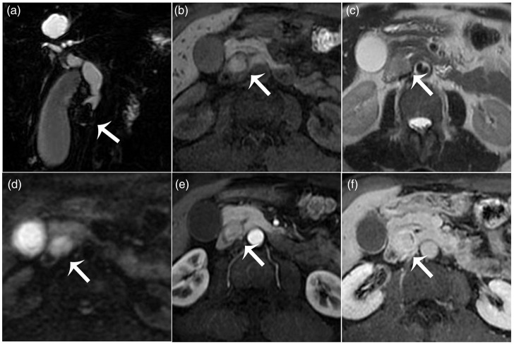
Primary biliary melanoma arises from proliferating melanocytes in the mucosal surface of the bile duct and is extremely rare. Since the vast majority of biliary melanomas represent metastases of cutaneous origin, accurate preoperative diagnosis of melanoma and exclusion of other primary sources are vital in cases involving primary lesions. Although melanomas with pigmented cells have typical signal characteristics, obtaining a non-invasive pre-treatment diagnosis remains difficult, due to their low incidence. Here, the case of a 61-year-old male Asian patient who presented with upper quadrant abdominal pain, swelling and jaundice for 2 weeks, and who was diagnosed with primary biliary melanoma following extensive preoperative blood analyses, computed tomography (CT) and magnetic resonance imaging (MRI), is described. Post-resection immunohistochemistry confirmed the diagnosis and the patient received six chemotherapy cycles of temozolomide and cisplatin, however, progression of multiple liver metastases was observed at the 18-month follow-up CT. The patient continued with pembrolizumab and died 17 months later. The present case of primary biliary melanoma is the first reported diagnosis based on typical MRI features and the exhaustive exclusion of a separate primary origin.
Keywords: Bile duct; imaging features.; immunohistochemistry; jaundice; magnetic resonance imaging; malignant melanoma.
Conflict of interest statement
The authors declare that there is no conflict of interest.
Figures


Similar articles
-
Primary hepatic melanoma: A case report of computed tomography and magnetic resonance imaging findings.Medicine (Baltimore). 2019 Jun;98(25):e16165. doi: 10.1097/MD.0000000000016165. Medicine (Baltimore). 2019. PMID: 31232974 Free PMC article.
-
Primary biliary tract melanoma: Report of a case and review of the literature.Int J Surg Case Rep. 2012;3(9):441-4. doi: 10.1016/j.ijscr.2012.05.008. Epub 2012 May 24. Int J Surg Case Rep. 2012. PMID: 22706296 Free PMC article.
-
Complete remission of the liver metastases of anorectal malignant melanoma with regional chemotherapy: a case report.Hepatogastroenterology. 2000 May-Jun;47(33):612-4. Hepatogastroenterology. 2000. PMID: 10918997
-
Pigmented villous nodular synovitis mimicking metastases on 18F-FDG PET/CT in a patient with rectal mucosal melanoma: a case report.BMC Musculoskelet Disord. 2020 Jan 8;21(1):13. doi: 10.1186/s12891-019-3034-x. BMC Musculoskelet Disord. 2020. PMID: 31914975 Free PMC article. Review.
-
Primary Malignant Melanoma in the Pineal Region: Case Report and Literature Review.J Neurol Surg A Cent Eur Neurosurg. 2018 Jul;79(4):344-352. doi: 10.1055/s-0038-1639504. Epub 2018 Apr 12. J Neurol Surg A Cent Eur Neurosurg. 2018. PMID: 29649851 Review.
References
-
- Tapia Rico G, Yong CH, Herrera Gómez RG. Adjuvant systemic treatment for high-risk resected non-cutaneous melanomas: what is the evidence? Crit Rev Oncol Hematol 2021; 167: 103503. - PubMed
-
- Zou Z, Ou Q, Ren Y, et al.. Distinct genomic traits of acral and mucosal melanomas revealed by targeted mutational profiling. Pigment Cell Melanoma Res 2020; 33: 601–611. - PubMed
-
- Yde SS, Sjoegren P, Heje M, et al.. Mucosal melanoma: a literature review. Curr Oncol Rep 2018; 20: 28. - PubMed
-
- Spencer KR, Mehnert JM. Mucosal melanoma: epidemiology, biology and treatment. Cancer Treat Res 2016; 167: 295–320. - PubMed
Publication types
MeSH terms
LinkOut - more resources
Full Text Sources
Medical
