. 2023 May;12(10):11983-11999.
doi: 10.1002/cam4.5873. Epub 2023 Mar 31.

ITGA5 promotes tumor angiogenesis in cervical cancer

Affiliations

ITGA5 promotes tumor angiogenesis in cervical cancer

Xiaohan Xu et al. Cancer Med. 2023 May.

Abstract

Purpose: Integrins are critical to cancer progression. Integrin alpha 5 (ITGA5) is correlated with the prognosis of cervical cancer patients. However, whether ITGA5 plays an active role in cervical cancer progression or not remains unknown.

Methods: ITGA5 protein expression was detected in 155 human cervical cancer tissues by immunohistochemistry. Data from The Cancer Genome Atlas were utilized to identify risk factors for the overall survival of cervical cancer patients and ITGA5-associated differentially expressed genes. Analyses of single-cell RNA-seq based on Gene Expression Omnibus datasets were performed to show the coexpression of ITGA5 and angiogenesis factors. Tube formation assay, 3D spheroid sprout assay, qRT-PCR, Western Blotting, ELISA, and immunofluorescence were conducted to explore the angiogenic function of ITGA5 in vitro and underlying mechanisms.

Results: High ITGA5 level was significantly correlated with increased risk in terms of overall survival and advanced disease stage in cervical cancer patients. ITGA5-associated differentially expressed genes linked ITGA5 to angiogenesis, and immunohistochemistry showed a positive correlation between ITGA5 and microvascular density in cervical cancer tissues. Moreover, tumor cells transfected with ITGA5-targeting siRNA decreased ability to promote endothelial tube formation in vitro. ITGA5/VEGFA coexpression was observed in a tumor cell subpopulation and the decreased endothelial angiogenesis by downregulating ITGA5 could be reversed by VEGFA. Bioinformatics analysis highlighted the PI3K-Akt signaling pathway as downstream of ITGA5. Downregulation of ITGA5 in tumor cells significantly decreased p-AKT and VEGFA levels. Fibronectin (FN1) coated cells or transfected with FN1-targeting siRNA showed fibronectin may play a critical role on ITGA5-mediated angiogenesis.

Conclusion: ITGA5 promotes angiogenesis and possibly be a potential predictive biomarker for poor survival of patients in cervical cancer.

Keywords: ITGA5; VEGFA; angiogenesis; cervical cancer.


Conflict of interest statement

The authors declare that there is no conflict of interest.

Figures

FIGURE 1
FIGURE 1
High ITGA5 expression is associated with poorer prognosis in cervical cancer patients. (A) Forest plots of univariate Cox regression analyses involving integrin superfamily members in overall survival (OS) of 306 cervical cancer patients in TCGA dataset. (B) Kaplan–Meier curves of OS of cervical cancer patients with ITGA5 high versus ITGA5 low tumors using GEPIA2 data based on TCGA (N = 292). (C) Kaplan–Meier curves of OS of cervical cancer patients with ITGA5 high versus ITGA5 low tumors using Kaplan–Meier plotter data based on TCGA (N = 304). (D) The expression of integrins in cervical cancer patients with different response to concurrent chemoradiotherapy in GSE168009 (NDB, no durable benefit; DCB, durable clinical benefit). Bar, SD; *p < 0.05, Student's t‐test. (E) Representative images of immunohistochemistry (IHC) staining for ITGA5 in cervical cancer tissues. Scale bar, 50 μm. (F) Kaplan–Meier curves of OS in cervical cancer patients stratified by ITGA5 IHC score. (A score greater than 6 as high ITGA5 expression; otherwise, low ITGA5 expression was noted).
FIGURE 2
FIGURE 2
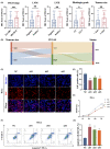
ITGA5 promotes progression of cervical cancer. (A) Scatter diagrams showing the distribution of ITGA5 IHC score in tumors grouped by FIGO stage, lymph node metastasis (LNM), lymph vascular space invasion (LVSI), histologic grade (G1, well differentiated tumor; G2‐3, moderate and poorly differentiated tumor), and tumor size. Bar, SD; Mann–Whitney U test. (B) The relationships among tumor size, level of ITGA5, and status of patients in the Sankey diagram. (C) Representative images of EdU assay of HeLa cells transfected with siITGA5 or negative control siRNA (NC). Scale bar, 50 μm. (D) The histogram shows the quantification results of the EdU assays of HeLa cells of three independent experiments. Bar, SD; One‐way ANOVA. (E) Cell proliferation curves generated with results of CCK‐8 assays of HeLa cells transfected with siITGA5 or NC of three independent experiments. One‐way ANOVA. (F) Representative images of flow cytometry apoptosis analysis of HeLa cells transfected with siITGA5 or NC. (G) The histogram shows the proportion of apoptotic HeLa cells (early apoptosis Q3 plus late apoptosis Q2) of three independent experiments. Bar, SD; One‐way ANOVA. *p < 0.05; **p < 0.01; ***p < 0.001; ****p < 0.0001.
FIGURE 3
FIGURE 3
ITGA5 is correlated with angiogenesis in cervical cancer. (A) TCGA database volcano plot demonstrates differentially expressed genes (DEGs) between cervical cancer patients with different ITGA5 expression (TCGA code CESC). (B) GSEA indicate that angiogenesis is significantly enriched in highly expressed ITGA5 associated DEGs. (C) The correlations between ITGA5 and angiogenesis pathway. Spearman correlation test. (D) Representative cases with high (NO.1) and low levels (NO.50) of ITGA5 that are correspondingly stained CD31 to identify microvessels are shown. Scale bar, 50 μm. (E) Correlation analysis of the immunohistochemistry score of ITGA5 and the microvessel density (MVD) in cervical cancer tissues (N = 57). Pearson correlation test. (F) Representative images of tube formation assays and 3D spheroid sprouting assays of HUVECs treated with conditional medium from HeLa cells transfected with siITGA5 or negative control siRNA (NC). The representative sprout is marked by green line. The histograms show the number of nodes and junctions of the tube formation assay and the average sprout length of 3D spheroid sprouting assay of three independent experiments. Scale bar, 50 μm. Bar, SD; One‐way ANOVA. *p < 0.05; **p < 0.01; ***p < 0.001.
FIGURE 4
FIGURE 4
ITGA5 promotes angiogenesis in vitro by regulating VEGFA. (A) Correlation analysis between ITGA5 expression and angiogenesis gene markers ANGPT1, ANGPT2, and VEGFA in cervical cancer by GEPIA2 based on TCGA dataset. Pearson correlation test. (B) qRT‐PCR for the mRNA level of ANGPT1, ANGPT2, VEGFA of HeLa and SiHa cells transfected with siITGA5 or negative control siRNA (NC). Three independent experiments. Bar, SD; One‐way ANOVA. (C) Western Blotting assay for the protein expression of ANGPT1, ANGPT2, VEGFA of HeLa and SiHa cells transfected with siITGA5 or NC. (D) Single‐cell RNA‐seq data of cell‐type and molecular subtype assignment of GSE168652, UMAP of cells from tumor tissue of cervical cancer patients, colored by clustering results. (E) Feature plots of relevant marker genes in immune cells (PTPRC) and cancer cells (CDH1, CDKN2A, and EPCAM) and the expression of ITGA5, ANGPT1, ANGPT2, and VEGFA in clusters of cancer cells. (F) Correlation analysis between ITGA5 and VEGFA expression of cervical cancer cells in single‐cell RNA‐seq data of GSE168652. Pearson correlation test. (G) ELISA assay for the level of VEGFA in conditional medium from cervical cancer cells transfected with siITGA5 or NC of three independent experiments. Bar, SD; One‐way ANOVA. (H) Representative images of tube formation assay and 3D spheroid sprouting assay of HUVECs stimulated with NC conditional medium, NC conditional medium + Bevacizumab, siITGA5 conditional medium, and siITGA5 conditional medium + VEGFA in HeLa cells. The representative sprout is marked by green line. The histograms show the number of nodes and junctions of the tube formation assay and the average sprout length of 3D spheroid sprouting assay of three independent experiments. Scale bar, 50 μm. Bar, SD; One‐way ANOVA; *p < 0.05; **p < 0.01; ***p < 0.001; ****p < 0.0001; ns, not significant.
FIGURE 5
FIGURE 5
ITGA5 regulates the AKT/VEGFA signaling pathway. (A) KEGG enriched pathway analysis in ITGA5‐associated differentially expressed genes from TCGA. (B) UMAP of ITGA5‐positive and ITGA5‐negative cells from tumor tissue of cervical cancer patients in GSE168652, colored by clustering results. (C) KEGG enriched pathway analysis in ITGA5‐positive cluster of GSE168652. (D) Western Blotting assay for the expression of AKT and p‐AKT of HeLa and SiHa cells transfected with siITGA5 or negative control siRNA (NC). (E) Western Blotting assay show the expression ITGA5, AKT, p‐AKT, and VEGFA in HeLa and SiHa cells treated with DMSO, 10 μM, and 15 μM MK‐2206 2HCI (AKT inhibitor).
FIGURE 6
FIGURE 6
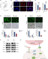
Fibronectin plays critical role in ITGA5‐mediated angiogenesis in vitro. (A) Correlation analysis between ITGA5 expression and FN1 in cervical cancer by GEPIA2 based on TCGA. Pearson correlation test. (B) Representative images of immunofluorescence staining of FN1 in HeLa cells with fibronectin substrate coated (+fibronectin) or control. Scale bar, 50 μm. (C) qRT‐PCR for the mRNA level of FN1 of HeLa and SiHa cells coated with fibronectin substrate (+fibronectin) or control. Four independent experiments. Bar, SD; Student's t‐test. (D, E) Representative images of tube formation assay and 3D spheroid sprouting assay of HUVECs stimulated with conditional medium form HeLa cells with fibronectin substrate coated (+fibronectin) or control and conditional medium from HeLa cells transfected with siFN1 or negative control siRNA (NC), respectively. Scale bar, 50 μm. The representative sprout is marked by green line. The histograms show the number of nodes and junctions of the tube formation assay and the average sprout length of 3D spheroid sprouting assay of three independent experiments. Bar, SD; Student's t‐test. (F) Western Blotting assay show the expression of FN1, AKT, p‐AKT, and VEGFA in HeLa and SiHa cells transfected with siFN1 or NC. (G) The schematic figure shows that ITGA5 promotes angiogenesis in cervical cancer by AKT/VEGFA axis and Fibronectin plays critical role in this pathway. *p < 0.05; ***p < 0.001; ****p < 0.0001.

Similar articles

Cited by

References

    1. Siegel RL, Miller KD, Fuchs HE, Jemal A. Cancer statistics, 2022. CA Cancer J Clin. 2022;72(1):7‐33. - PubMed
    1. Castle PE, Einstein MH, Sahasrabuddhe VV. Cervical cancer prevention and control in women living with human immunodeficiency virus. CA Cancer J Clin. 2021;71(6):505‐526. - PMC - PubMed
    1. Attademo L, Tuninetti V, Pisano C, et al. Immunotherapy in cervix cancer. Cancer Treat Rev. 2020;90:102088. - PubMed
    1. Desgrosellier JS, Cheresh DA. Integrins in cancer: biological implications and therapeutic opportunities. Nat Rev Cancer. 2010;10(1):9‐22. - PMC - PubMed
    1. Kuninty PR, Bansal R, De Geus SWL, et al. ITGA5 inhibition in pancreatic stellate cells attenuates desmoplasia and potentiates efficacy of chemotherapy in pancreatic cancer. Sci Adv. 2019;5(9):eaax2770. - PMC - PubMed

Publication types

MeSH terms