. 2023 Jun;145(6):793-814.
doi: 10.1007/s00401-023-02565-1. Epub 2023 Mar 31.

C-terminal frameshift variant of TDP-43 with pronounced aggregation-propensity causes rimmed vacuole myopathy but not ALS/FTD

Affiliations

C-terminal frameshift variant of TDP-43 with pronounced aggregation-propensity causes rimmed vacuole myopathy but not ALS/FTD

Pedro Ervilha Pereira et al. Acta Neuropathol. 2023 Jun.

Abstract

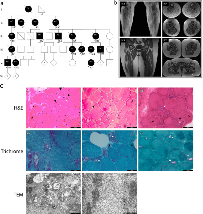
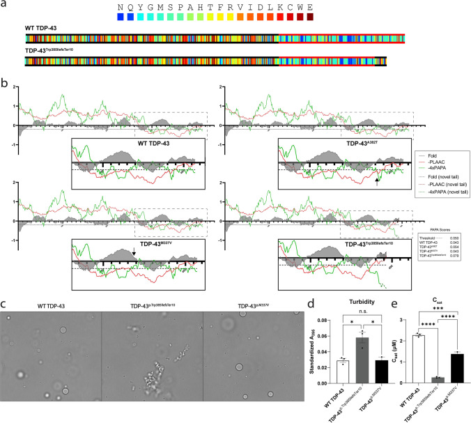
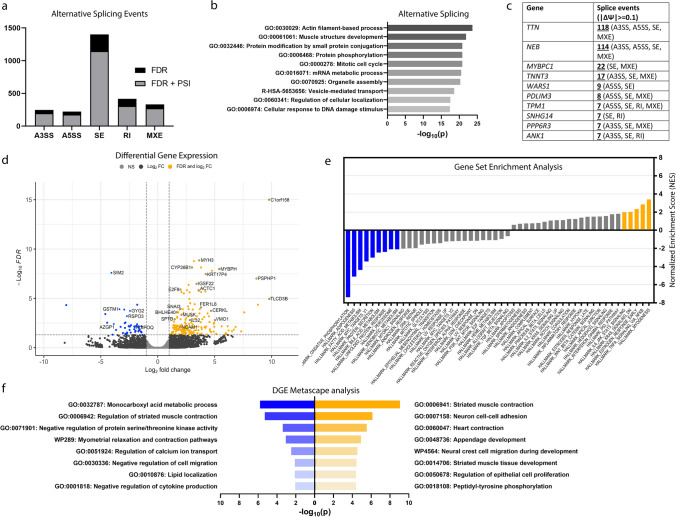
Neuronal TDP-43-positive inclusions are neuropathological hallmark lesions in frontotemporal dementia (FTD) and amyotrophic lateral sclerosis (ALS). Pathogenic missense variants in TARDBP, the gene encoding TDP-43, can cause ALS and cluster in the C-terminal prion-like domain (PrLD), where they modulate the liquid condensation and aggregation properties of the protein. TDP-43-positive inclusions are also found in rimmed vacuole myopathies, including sporadic inclusion body myositis, but myopathy-causing TDP-43 variants have not been reported. Using genome-wide linkage analysis and whole exome sequencing in an extended five-generation family with an autosomal dominant rimmed vacuole myopathy, we identified a conclusively linked frameshift mutation in TDP-43 producing a C-terminally altered PrLD (TDP-43p.Trp385IlefsTer10) (maximum multipoint LOD-score 3.61). Patient-derived muscle biopsies showed TDP-43-positive sarcoplasmic inclusions, accumulation of autophagosomes and transcriptomes with abnormally spliced sarcomeric genes (including TTN and NEB) and increased expression of muscle regeneration genes. In vitro phase separation assays demonstrated that TDP-43Trp385IlefsTer10 does not form liquid-like condensates and readily forms solid-like fibrils indicating increased aggregation propensity compared to wild-type TDP-43. In Drosophila TDP-43p.Trp385IlefsTer10 behaved as a partial loss-of-function allele as it was able to rescue the TBPH (fly ortholog of TARDBP) neurodevelopmental lethal null phenotype while showing strongly reduced toxic gain-of-function properties upon overexpression. Accordingly, TDP-43p.Trp385IlefsTer10 showed reduced toxicity in a primary rat neuron disease model. Together, these genetic, pathological, in vitro and in vivo results demonstrate that TDP-43p.Trp385IlefsTer10 is an aggregation-prone partial loss-of-function variant that causes autosomal dominant vacuolar myopathy but not ALS/FTD. Our study genetically links TDP-43 proteinopathy to myodegeneration, and reveals a tissue-specific role of the PrLD in directing pathology.

Keywords: ALS/FTD; Drosophila; Genetics; Myopathy; Phase separation; TDP-43.


Conflict of interest statement

J.S. is a consultant for Dewpoint Therapeutics, Confluence Therapeutics, ADRx, and Neumora. The other authors have nothing to declare.

Figures

Fig. 1
Fig. 1
Identification of TDP-43p.Trp385IlefsTer10 as a cause of autosomal dominant rimmed vacuole myopathy. a Pedigree of the family in which the TDP-43p.Trp385ilefsTer10 variant was identified. Individuals identified with “m/- “ are affected individuals that have either been genotyped or in which the genotype was reconstructed based on segregation. Individuals that were genotyped are identified with an asterisk (*). b Coronal and axial plane T1-weighted MRI images of the lower limb muscles of patients IV.3 and IV.4. Fatty atrophy of the muscles in the anterior, lateral and posterior compartments of the distal lower extremities in patient IV.4 (top left). Global muscular atrophy of the pelvic girdle and proximal lower extremities in patient IV.3 (bottom left). Fatty atrophy of the gluteus maximus and tensor fasciae latae (patient IV.3, bottom right, white arrowheads). Bilateral focal infiltration in the vastus lateralis of the quadriceps muscles (patients IV.3 and IV.4, white arrows). c Histological images of patient muscle biopsies: i-iii. H&E of tissue from patients IV.5, IV.4 and III.5, respectively. Tissue sections show signs of myopathy with an uneven distribution of fibre size, presence of centralized nuclei (black arrowheads) and increased fibrotic tissue proliferation (white arrows). Scale bar = 100 μm. iv-vi. Modified Gomori trichrome staining of muscle tissue from patients IV.5, IV.4 and III.1, respectively. Staining shows the presence of rimmed vacuoles (white arrows), along with infiltration of fatty tissue (black arrowheads). Scale bar = 50 μm. vii. TEM of patient III.5 muscle reveals signs of enhanced autophagy with the presence of multiple autophagic vesicles (black arrow) and viii. the accumulation of fibrous material (black arrowhead)
Fig. 2
Fig. 2
TDP-43p.Trp385IlefsTer10 protein is expressed in mutant muscle tissue and fibroblasts. a LOD-score plot of chromosome 1 showing conclusive linkage (Max. multipoint LOD Score = 3.61) to chromosome 1p36.22 at 8.1–13.8 cM along with the variants found in the region of interest. Further information on the classification of the variants can be found in Supplementary table 1. b Graphical representation of the structure of the TDP-43 protein and the changes to the protein sequence caused by TDP-43p.Trp385IlefsTer10. Phosphorylatable sites Ser409 and Ser410 (represented with a “P”) are lost in the mutant protein, meaning this phosphorylation marker is lost in TDP-43p.Trp385IlefsTer10. c TDP-43 levels in muscle tissue and fibroblasts collected from patients. Muscle of patients shows a clear reduction in TDP-43 levels, whereas TDP-43 levels in fibroblasts are evenly distributed between the wild-type (green arrowhead) and mutant (red arrowhead) forms of TDP-43. d Relative quantification of the levels of WT TDP-43 protein in patient tissues versus control (npatient = 2, ncontrol = 3). Protein levels were normalised against GAPDH levels for all conditions. e Despite the altered levels of TDP-43 in muscle, the levels of phosphorylated TDP-43 remain unaffected in both muscle and fibroblasts. f Relative quantification of the levels of phosphorylated TDP-43 protein in patient tissues versus control (npatient = 2, ncontrol = 3). Protein levels were normalised against GAPDH levels for all conditions. Statistical analysis performed using the standard two-way ANOVA test using multiple comparisons; *p <  = 0.05
Fig. 3
Fig. 3
Phosphorylation state of TDP-43p.Trp385IlefsTer10 accumulation suggests a predominantly mutant protein composition. Immunofluorescent staining of control, sIBM and patient muscle tissue. a TDP-43 shows primarily nuclear staining, with presence of TDP-43 accumulation in sIBM and Patient tissue (insets). b Staining for pTDP-43[Ser369] reveals that TDP-43 accumulations are positive for phosphorylation of Ser396 in both sIBM and TDP-43p.Trp385IlefsTer10 patient tissues. c This is not the case for pTDP-43[Ser409/410], where only sIBM tissues have TDP-43 accumulation positive for this phosphorylation marker. Scale bar = 50 μm
Fig. 4
Fig. 4
TDP-43p.Trp385IlefsTer10 is aggregation-prone compared to WT TDP-43 and TDP-43p.M337V. a-b PLAAC prediction for prion-like domains shows that the PrLD of TDP-43p.Trp385IlefsTer10, marked with red lines in the color-coded sequence visualizations (a), and adjusted PAPA and negative PLAAC scores (b), spans a smaller region when compared to wild-type TDP-43 and the two ALS mutants. The black lines in the color-coded sequence (a) indicate that the aa. frequencies correspond to the background frequencies and hence no PrLD is identified. Each aa. is color-coded by its enrichment log-likelihood ratio in PrLDs (blue = high, red = low). The default settings (Lcore = 60 and S. cerevisiae background frequencies) were used to run the PLAAC application. Despite the smaller length in PrLD, PAPA predictions (green lines in b), indicate a higher propensity for aggregative potential in the mutant variant. The dashed line in b marks the threshold of the PAPA-score for prion-like behaviour. Minimal differences in Fold Index (FI), a prediction to identify intrinsically unfolded protein regions (= negative score), are observed between wild type TDP43, ALS TDP-43 mutants and PrLD of TDP-43p.Trp385IlefsTer10. c-e WT TDP-43-MBP-His and TDP-43p.Trp385IlefsTer10-MBP-His and TDP-43p.M337V-MBP-His (5 µM) were incubated for 30 min at room temperature. c Representative brightfield microscopy images indicate that WT TDP-43 and TDP-43p.M337V form spherical droplets, whereas TDP-43p.Trp385IlefsTer10 forms irregularly shaped aggregates. The scale bar represents 10 µm. d Standardized turbidity, measured in a plate reader at 395 nm, is significantly higher for TDP-43p.Trp385IlefsTer10 versus WT TDP-43 and TDP-43p.M337V. Bars represent means ± SEM (n = 3), and each individual data point is shown. An ordinary one-way ANOVA with Tukey’s multiple comparisons test was performed, *p < 0.05. e The saturation concentration (Csat) of each sample was measured after centrifugation. Csat is significantly lower for TDP-43p.Trp385IlefsTer10 versus WT TDP-43 and TDP-43p.M337V. Bars represent means ± SEM (n = 3), and each individual data point is shown. Statistical analysis performed using an ordinary one-way ANOVA with Tukey’s multiple comparisons test, ***p < 0.001, ****p < 0.0001
Fig. 5
Fig. 5
TDP-43p.Trp385IlefsTer10 behaves as a partial loss-of-function allele with reduced toxicity in Drosophila. a Expression levels of human TDP-43 in control and transgenic fly lines using the pan-neuronal nSyb-Gal4 driver. b Rescue assay results showing the rate of offspring frequency in TBPH null flies rescued with the TDP-43 constructs of interest driven by the nSyb-Gal4 pan-neuronal driver (right) and the D42-Gal4 motor neuron-specific driver (left). c Combined survival assay performed on overexpression models of the TDP-43 variants of interest driven by the nSyb-Gal4 neuronal driver. d Combined climbing assay performed on transgenic models of the TDP-43 variants of interest driven by the nSyb-Gal4 pan-neuronal driver. Fraction of flies capable of completing the assay was plotted weekly throughout the course of the assay. e Expression levels of human TDP-43 in control and transgenic fly lines using the nSyb-Gal80 inducible driver and the tubulin-Gal80 inducible driver. For the inducible drivers, the levels of protein expression at 29 ℃ vs. 18 ℃ confirm that the system is reliably inducing higher levels of expression when flies are kept at a higher environmental temperature (29 ℃). f Combined survival results for both male and female transgenic and control flies with variant adult onset expression driven in a tissue-specific manner. From left to right: whole body expression with Tubulin-Gal80, muscular expression with Mef2-Gal80 and pan-neuronal expression with nSyb-Gal80. Expression of hTDP-43 variants was induced by moving the flies into a 29 ℃ environment. g Rodent mixed cortical primary neurons were transfected with the indicated constructs and tracked by automated fluorescence microscopy. Neuronal survival and time of death was assessed using objective criteria (described in Supplementary Fig. 4, online resource), and cumulative risk of death plotted for neurons in each condition. N = number of neurons, combined from 3 biological replicates. *hazard ratio (HR) = 1.29, p = 6.7 × 10–7; **HR = 1.31, p = 8.0 × 10–8; ***HR = 1.56, p < 2 × 10–16; #HR = 1.17, p = 0.003. HR and p value determined by cox proportional hazards analysis, stratifying among biological replicates. Statistical analysis performed using the one-way ANOVA test (Kruskal–Wallis test); *p ≤ 0.05, **p ≤ 0.01 (unless otherwise stated)
Fig. 6
Fig. 6
Abnormal splicing of sarcomeric genes, increased expression of muscle regeneration genes and decreased expression of mitochondrial and lipid metabolism genes in TDP-43p.Trp385IlefsTer10 muscle-derived transcriptomes. a Distribution of the number of alternative splicing events identified across each AS event type filtered by FDR ≤ 0.01 (FDR) and FDR ≤ 0.01 +|ΔΨ|≥ 0.1 (FDR + PSI). b Metascape analysis and top GO terms associated with the aggregated genes suffering AS events. c Top genes with multiple alternative splicing events identified and the respective types. Inclusion level difference filtered at |ΔΨ|≥ 0.1. d Volcano plot of log2[fold change] values against the respective FDR values. Cutoff values for FDR ≤ 0.05 and |log2FC|> 1. e Pathways identified by pre-ranked GSEA of the differentially expressed genes dataset and ranked by normalized enrichment score (NES). Pathways identified were considered significant at an FDR ≤ 0.01 (highlighted; blue—downregulated, gold—upregulated). f Metascape analysis of the top GO terms associated with the downregulated (blue) and upregulated (gold) genes identified in the DGE dataset

References

    1. Arai T, Hasegawa M, Akiyama H, Ikeda K, Nonaka T, Mori H, et al. TDP-43 is a component of ubiquitin-positive tau-negative inclusions in frontotemporal lobar degeneration and amyotrophic lateral sclerosis. Biochem Biophys Res Commun. 2006;351:602–611. doi: 10.1016/j.bbrc.2006.10.093. - DOI - PubMed
    1. Archbold HC, Jackson KL, Arora A, Weskamp K, Tank EM-H, Li X, et al. TDP43 nuclear export and neurodegeneration in models of amyotrophic lateral sclerosis and frontotemporal dementia. Sci Rep. 2018;8:4606. doi: 10.1038/s41598-018-22858-w. - DOI - PMC - PubMed
    1. Arrasate M, Mitra S, Schweitzer ES, Segal MR, Finkbeiner S. Inclusion body formation reduces levels of mutant huntingtin and the risk of neuronal death. Nature. 2004;431:805–810. doi: 10.1038/nature02998. - DOI - PubMed
    1. Arseni D, Hasegawa M, Murzin AG, Kametani F, Arai M, Yoshida M, et al. Structure of pathological TDP-43 filaments from ALS with FTLD. Nature. 2022;601:139–143. doi: 10.1038/s41586-021-04199-3. - DOI - PMC - PubMed
    1. Barmada SJ, Serio A, Arjun A, Bilican B, Daub A, Ando DM, et al. Autophagy induction enhances TDP43 turnover and survival in neuronal ALS models. Nat Chem Biol. 2014;10:677–685. doi: 10.1038/nchembio.1563. - DOI - PMC - PubMed

Publication types

Supplementary concepts