. 2023 Aug;149(10):7689-7701.
doi: 10.1007/s00432-023-04721-2. Epub 2023 Mar 31.

Mitotic phosphorylation of Tau/MAPT modulates cell cycle progression in prostate cancer cells

Affiliations

Mitotic phosphorylation of Tau/MAPT modulates cell cycle progression in prostate cancer cells

Letizia Clementi et al. J Cancer Res Clin Oncol. 2023 Aug.

Abstract

Purpose: Tau/MAPT (microtubule associated protein tau) protein is actively studied for the pathologic consequences of its aberrant proteostasis in central nervous system leading to neurodegenerative diseases. Besides its ability to generate insoluble toxic oligomers, Tau homeostasis has attracted attention for its involvement in the formation of the mitotic spindle. This evidence, in association with the description of Tau expression in extra-neuronal tissues, and mainly in cancer tissues, constitutes the rationale for a more in-depth investigation of Tau role also in neoplastic diseases.

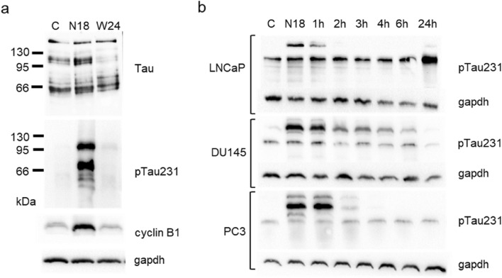
Methods: In our study, we investigated the expression of phosphorylated Tau in prostate cancer cell lines with particular focus on the residue Thr231 present in microtubule binding domain.

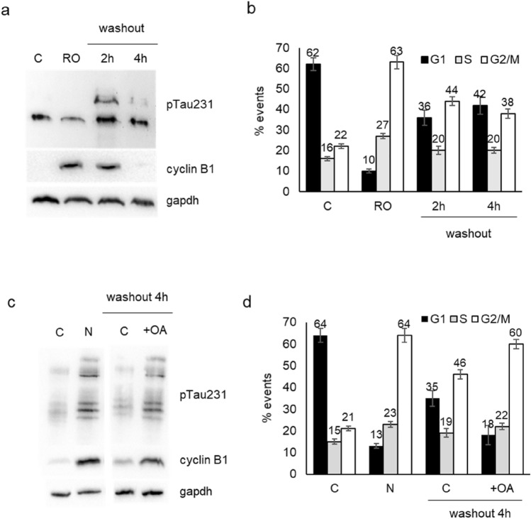
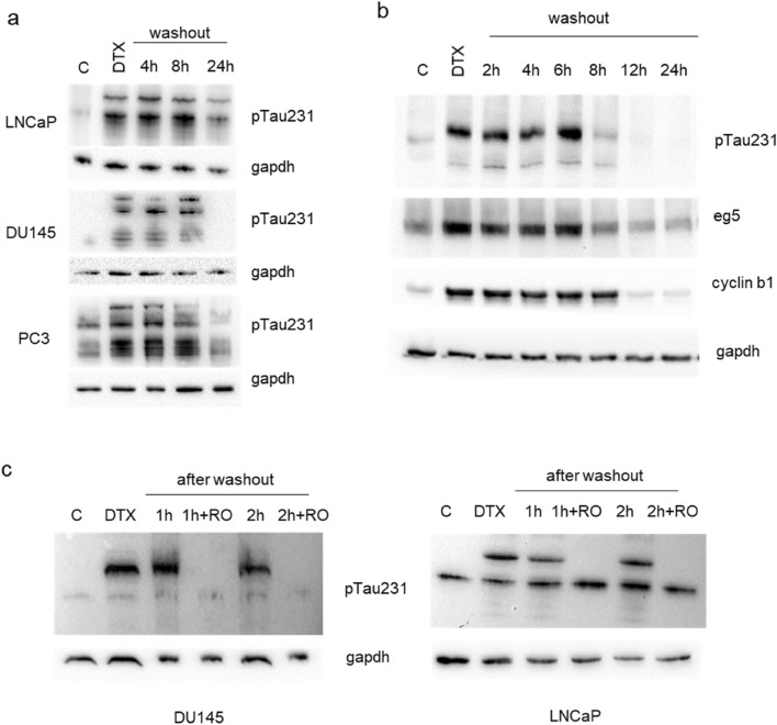
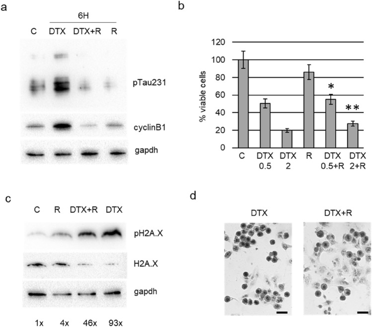
Results: The analysis of prostate cancer cells synchronized with nocodazole demonstrated that the expression of Tau protein phosphorylated at residue Thr231 is restricted to G2/M cell cycle phase. The phosphorylated form was unable to bind tubulin and it does not localize on mitotic spindle. As demonstrated by the use of specific inhibitors, the phosphorylation status of Tau is under the direct control of cdk5 and PP2A, while cdk1 activation was able to exert an indirect control. These mechanisms were also active in cells treated with docetaxel, where counteracting the expression of the dephosphorylated form, by kinase inhibition or protein silencing, determined resistance to drug toxicity.

Conclusions: We hypothesize that phosphorylation status of Tau is a key marker for G2/M phase in prostate cancer cells and that the forced modulation of Tau phosphorylation can interfere with the capacity of cell to efficiently progress through G2/M phase.

Keywords: Cancer therapy; Cell cycle; Docetaxel; Mitosis; Prostate cancer.


Conflict of interest statement

The authors declare that they have no conflict of interests.

Figures

Fig. 1
Fig. 1
Tau protein electrophoretic pattern visualized by western blot after cell cycle synchronization induced by nocodazole. a Total cell lysates from control DU145 cells (C), cells treated for 18 h with nocodazole (N18) and cells treated for 18 h with nocodazole and then washed out for further 24 h (W24), were analysed for the expression of total Tau, Tau phosphorylated at Thr231 (pTau231) and cyclin B1. b Total cell lysates from LNCaP, DU145 and PC3 cells were subjected to western blot analysis for the expression of Tau phosphorylated at Thr231 (pTau231). All cell lines were treated for 18 h with nocodazole and cell lysates were recovered at 18 h (N18) and in a time course after washing out (1 h, 2 h, 3 h, 4 h, 6 h, 24 h). For each western blot gapdh expression was showed as loading control
Fig. 2
Fig. 2
Analysis of the association between β-tubulin and Tau. a Total cell lysates from LNCaP, DU145 and PC3 cells were immunoprecipitated for β-tubulin and resulting pellet was probed with anti-Tau antibody (left). The supernatants recovered after immunoprecipitation (immunodepleted samples) were also subjected to western blot for Tau expression (right panel). b Representative images from immunofluorescence analysis of DU145 cells stained for the detection of β-tubulin (green), nuclei (blue) total Tau (red, upper row) or Tau phosphorylated at Thr231 (pTau231, red, lower row). The last image of each row is the digital overlay of the previous images (merge). The white bar is a scale of 20 μm
Fig. 3
Fig. 3
Expression of Tau phosphorylated at Thr231 and cell cycle progression in DU145 cells. a Western blot analysis for pTau231 and cyclin B1 in total cell lysates from control cells (C), cells treated for 18 h with RO-3306 (RO) and cells treated for 18 h with RO-3306, washed out and recovered after 2 h or 4 h. Gapdh expression in the same blot was visualized as loading control. b DU145 cells subjected to same protocol described in (a) were analysed by cytometry after propidium iodide staining and percentage of events in each phase of cell cycle is reported in the histogram. Data represent the mean of three independent experiments ± SD. c Western blot analysis for pTau231 and cyclin B1 in total cell lysates from control cells (C), cells treated for 18 h with nocodazole (N) and cells treated for 18 h with nocodazole, washed out, treated (+ OA) or not (C) with okadaic acid and recovered after 4 h. Gapdh expression in the same blot was visualized as loading control. d DU145 cells subjected to the same protocol described in (c) were analysed by cytometry for cell cycle analysis and percentage of events in each phase is reported in the histogram. Data represent the mean of three independent experiments ± SD
Fig. 4
Fig. 4
Expression of Tau phosphorylated at Thr231 in PCa cells treated with docetaxel. a LNCaP, DU145 and PC3 cells were treated with docetaxel and total cell lysates were recovered at 18 h (DTX) and in a time course after washout (4 h, 8 h, 24 h). For each western blot, gapdh expression was showed as loading control. b DU145 cells were treated with docetaxel and total cell lysates were recovered at 18 h (DTX) and in a time course after washout (2 h, 4 h, 6 h, 8 h, 12 h, 24 h). c Total cell lysates from DU145 and LNCaP cells were subjected to western blot analysis for the expression pTau231. Cell lines were treated with docetaxel and cell lysates were recovered at 18 h (DTX) and in a time course after washout (1 h and 2 h) in presence or not of RO-3306 (+ RO). For each western blot, gapdh expression was showed as loading control
Fig. 5
Fig. 5
Effect of roscovitine treatment in DU145 cells. a Cells were treated for 6 h with docetaxel, with docetaxel plus roscovitine (DTX + R) or with roscovitine (R) and total cell lysates were analysed by western blot for the expression of pTau231, cyclin B1 and gapdh. b Viable cells were measured by trypan blue exclusion test after treatment for 24 h with docetaxel 0.5 nM (DTX 0.5), docetaxel 2 nM (DTX 2), roscovitine (R), docetaxel 0.5 nM plus roscovitine (DTX 0.5 + R), docetaxel 2 nM plus roscovitine (DTX 2 + R). Data are represented as percentage considering control (C) as 100%. Results are the mean of three independent experiments ± SD. *p < 0.05 respect DTX 0.5; **p < 0.05 respect DTX 0.5. c Cells were treated for 24 h with docetaxel (DTX), with docetaxel plus roscovitine (DTX + R) or with roscovitine (R) and total cell lysates were analysed by western blot for the expression of pH2A.X, H2A.X and gapdh. Densitometric analysis of each pH2A.X band was performed normalizing for its respective H2A.X and gapdh bands and resulting values are showed. d Representative images of DU145 cells stained with haematoxylin after treatment for 24 h with 2 nM docetaxel (DTX) or with 2 nM docetaxel plus roscovitine (DTX + R). The black bar is a scale of 20 μm
Fig. 6
Fig. 6
Effect of Tau silencing in DU145 cells. a DU145 cells were transfected with siRNA targeting Tau (silTau) or with scrambled siRNA (C), and after 24 h were treated with docetaxel for further 24 h. At the end point cell lysates were analyzed for pTau231, cyclin B1 and gapdh expression. Densitometry of cyclin B1 bands respect to gapdh bands was performed and resulting values are indicated below cyclin B1 blot. b DU145 cells were transfected with siRNA targeting Tau (silTau) or with scrambled siRNA (C), and after 24 h were treated with docetaxel for further 24 h. At the end point cell lysates were analyzed for pTau231, parp1, cyclin B1 and gapdh expression. c DU145 cells were subjected to the same protocol described in (a) and analysed by cytometry after propidium iodide staining. Percentage of events ± SD for each cell cycle phase and in the sub-G1 region are showed in the table, and representative profiles acquired in logarithmic mode are showed in the upper panels, evidencing the sub-G1 region. Data are means of three independent experiments ± SD. d, e Expression levels, reported as log2 values, of MAPT in DU145 and PC3 prostate cancer cell lines, sensitive (original cell line) and resistant (R) to docetaxel (DTX): boxplot showing MAPT expression reported in the GSE33455 dataset (d); boxplot showing MAPT expression reported in the GSE158494 dataset (e). Statistical significance was calculated by using GEO2R software

References

    1. Alonso AD, Di Clerico J, Li B, Corbo CP, Alaniz ME, Grundke-Iqbal I, Iqbal K (2010) Phosphorylation of Tau at Thr212, Thr231, and Ser262 combined causes neurodegeneration. J Biol Chem 285:30851–30860 - PMC - PubMed
    1. Alquezar C, Arya S, Kao AW (2020) Tau post-translational modifications: dynamic transformers of tau function, degradation, and aggregation. Front Neurol 11:595532 - PMC - PubMed
    1. Andorfer C, Acker CM, Kress Y, Hof PR, Duff K, Davies P (2005) Cell-cycle reentry and cell death in transgenic mice expressing nonmutant human Tau isoforms. J Neurosci 25:5446–5454 - PMC - PubMed
    1. Andreadis A (2005) Tau gene alternative splicing: expression patterns, regulation and modulation of function in normal brain and neurodegenerative diseases. Biochem Biophys Acta 1739:91–103 - PubMed
    1. Asada-Utsugi M, Uemura K, Ayaki T, Minamiyama S, Hikiami R, Morimura T, Shodai A, Ueki T, Takahashi R, Kinoshita A, Urushitani M (2022) Failure of DNA double-strand break repair by Tau mediates Alzheimer’s disease pathology in vitro. Commun Biol 5:358 - PMC - PubMed