

Binding of Venezuelan Equine Encephalitis Virus Inhibitors to Importin-α Receptors Explored with All-Atom Replica Exchange Molecular Dynamics
- PMID: 37001021
- PMCID: PMC10358320
- DOI: 10.1021/acs.jpcb.3c00429
Binding of Venezuelan Equine Encephalitis Virus Inhibitors to Importin-α Receptors Explored with All-Atom Replica Exchange Molecular Dynamics
Abstract
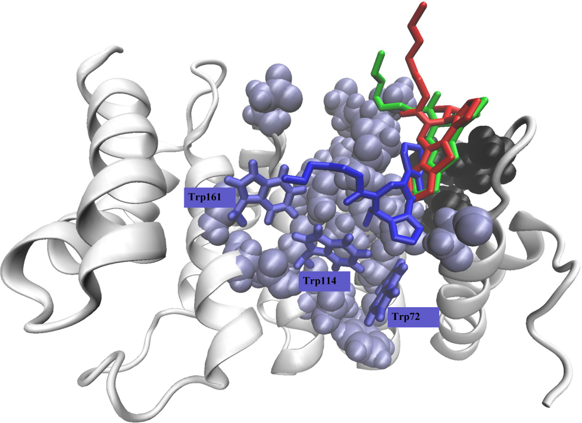
Although Venezuelan equine encephalitis virus (VEEV) is a life-threatening pathogen with a capacity for epidemic outbreaks, there are no FDA-approved VEEV antivirals for humans. VEEV cytotoxicity is partially attributed to the formation of a tetrameric complex between the VEEV capsid protein, the nuclear import proteins importin-α and importin-β, and the nuclear export protein CRM1, which together block trafficking through the nuclear pore complex. Experimental studies have identified small molecules from the CL6662 scaffold as potential inhibitors of the viral nuclear localization signal (NLS) sequence binding to importin-α. However, little is known about the molecular mechanism of CL6662 inhibition. To address this issue, we employed all-atom replica exchange molecular dynamics simulations to probe, in atomistic detail, the binding mechanism of CL6662 ligands to importin-α. Three ligands, including G281-1485 and two congeners with varying hydrophobicities, were considered. We investigated the distribution of ligand binding poses, their locations, and ligand specificities measured by the strength of binding interactions. We found that G281-1485 binds nonspecifically without forming well-defined binding poses throughout the NLS binding site. Binding of the less hydrophobic congener becomes strongly on-target with respect to the NLS binding site but remains nonspecific. However, a more hydrophobic congener is a strongly specific binder and the only ligand out of three to form a well-defined binding pose, while partially overlapping with the NLS binding site. On the basis of free energy estimates, we argue that all three ligands weakly compete with the viral NLS sequence for binding to importin-α in an apparent compromise to preserve host NLS binding. We further show that all-atom replica exchange binding simulations are a viable tool for studying ligands binding nonspecifically without forming well-defined binding poses.
Conflict of interest statement
Notes: The authors declare no con ict of interest.
Figures


Similar articles
-
Competitive Binding of Viral Nuclear Localization Signal Peptide and Inhibitor Ligands to Importin-α Nuclear Transport Protein.J Chem Inf Model. 2024 Jul 8;64(13):5262-5272. doi: 10.1021/acs.jcim.4c00626. Epub 2024 Jun 13. J Chem Inf Model. 2024. PMID: 38869471 Free PMC article.
-
Binding of Inhibitors to Nuclear Localization Signal Peptide from Venezuelan Equine Encephalitis Virus Capsid Protein Explored with All-Atom Replica Exchange Molecular Dynamics.ACS Omega. 2024 Sep 16;9(38):40259-40268. doi: 10.1021/acsomega.4c06981. eCollection 2024 Sep 24. ACS Omega. 2024. PMID: 39346821 Free PMC article.
-
Binding of viral nuclear localization signal peptides to importin-α nuclear transport protein.Biophys J. 2023 Sep 5;122(17):3476-3488. doi: 10.1016/j.bpj.2023.07.024. Epub 2023 Aug 4. Biophys J. 2023. PMID: 37542371 Free PMC article.
-
Nuclear import and export inhibitors alter capsid protein distribution in mammalian cells and reduce Venezuelan Equine Encephalitis Virus replication.Antiviral Res. 2013 Dec;100(3):662-72. doi: 10.1016/j.antiviral.2013.10.004. Epub 2013 Oct 22. Antiviral Res. 2013. PMID: 24161512
-
Venezuelan Equine Encephalitis Virus Capsid-The Clever Caper.Viruses. 2017 Sep 29;9(10):279. doi: 10.3390/v9100279. Viruses. 2017. PMID: 28961161 Free PMC article. Review.
Cited by
-
Competitive Binding of Viral Nuclear Localization Signal Peptide and Inhibitor Ligands to Importin-α Nuclear Transport Protein.J Chem Inf Model. 2024 Jul 8;64(13):5262-5272. doi: 10.1021/acs.jcim.4c00626. Epub 2024 Jun 13. J Chem Inf Model. 2024. PMID: 38869471 Free PMC article.
-
Binding of Inhibitors to Nuclear Localization Signal Peptide from Venezuelan Equine Encephalitis Virus Capsid Protein Explored with All-Atom Replica Exchange Molecular Dynamics.ACS Omega. 2024 Sep 16;9(38):40259-40268. doi: 10.1021/acsomega.4c06981. eCollection 2024 Sep 24. ACS Omega. 2024. PMID: 39346821 Free PMC article.
-
Can Free Energy Perturbation Simulations Coupled with Replica-Exchange Molecular Dynamics Study Ligands with Distributed Binding Sites?J Chem Inf Model. 2023 Aug 14;63(15):4791-4802. doi: 10.1021/acs.jcim.3c00631. Epub 2023 Aug 2. J Chem Inf Model. 2023. PMID: 37531558 Free PMC article.
References
Publication types
MeSH terms
Substances
Grants and funding
LinkOut - more resources
Full Text Sources
