Review
. 2023 Jun 20:92:299-332.
doi: 10.1146/annurev-biochem-032620-104401. Epub 2023 Mar 31.

Mitochondrial DNA Release in Innate Immune Signaling

Affiliations
Review

Mitochondrial DNA Release in Innate Immune Signaling

Laura E Newman et al. Annu Rev Biochem. .

Abstract

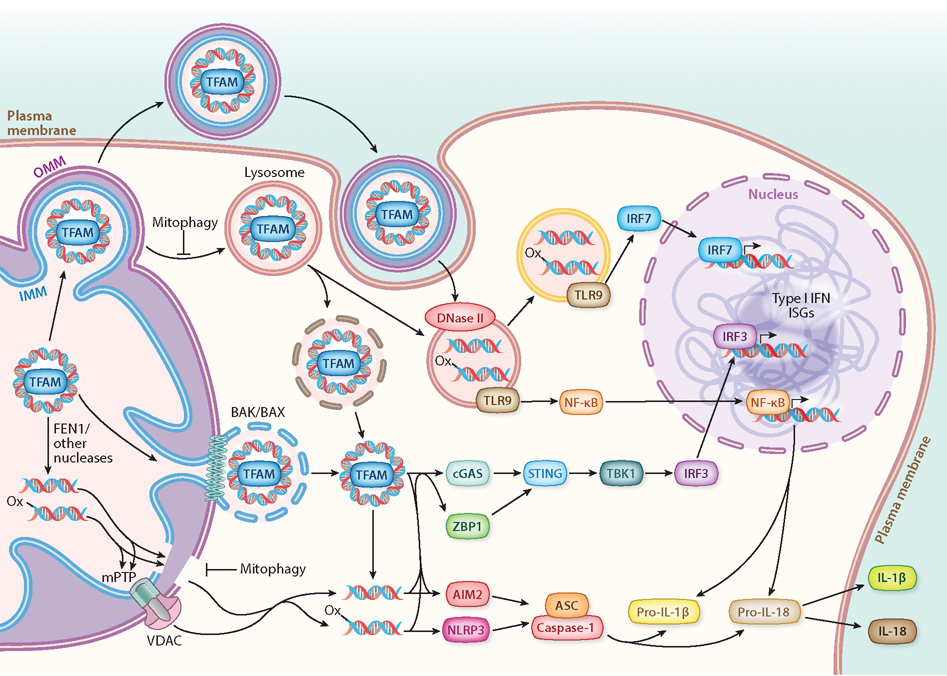
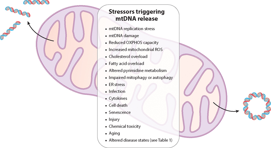
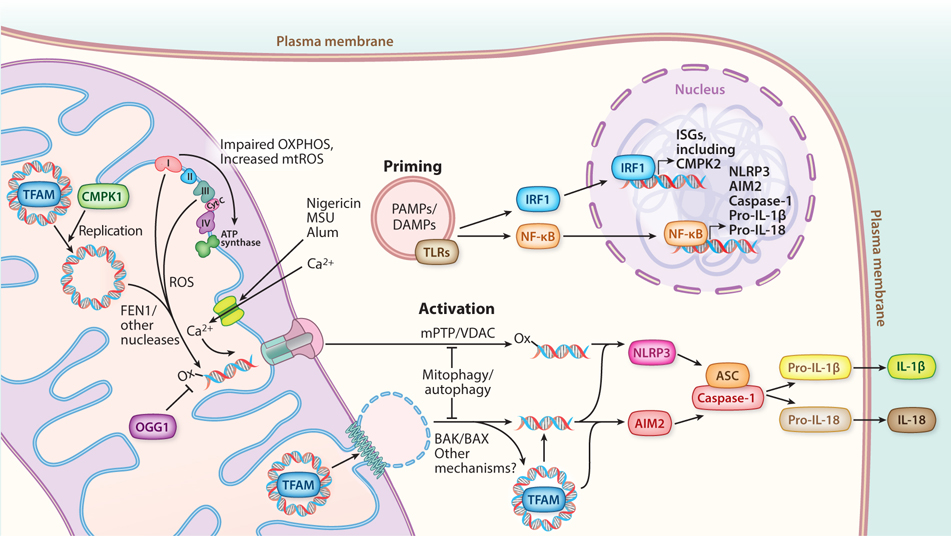
According to the endosymbiotic theory, most of the DNA of the original bacterial endosymbiont has been lost or transferred to the nucleus, leaving a much smaller (∼16 kb in mammals), circular molecule that is the present-day mitochondrial DNA (mtDNA). The ability of mtDNA to escape mitochondria and integrate into the nuclear genome was discovered in budding yeast, along with genes that regulate this process. Mitochondria have emerged as key regulators of innate immunity, and it is now recognized that mtDNA released into the cytoplasm, outside of the cell, or into circulation activates multiple innate immune signaling pathways. Here, we first review the mechanisms through which mtDNA is released into the cytoplasm, including several inducible mitochondrial pores and defective mitophagy or autophagy. Next, we cover how the different forms of released mtDNA activate specific innate immune nucleic acid sensors and inflammasomes. Finally, we discuss how intracellular and extracellular mtDNA release, including circulating cell-free mtDNA that promotes systemic inflammation, are implicated in human diseases, bacterial and viral infections, senescence and aging.

Keywords: DNA sensing; disease; inflammation; mitochondria; pore.


Figures

Figure 1
Figure 1
mtDNA-driven innate immune signaling. Release of mtDNA can occur through several different mechanisms, including IMM herniation through BAK/BAX pores in the OMM or escape involving mPTP and VDAC pores. Escape of mtDNA may also occur through leaky mitochondria or lysosomes, arising from defective mitophagy, autophagy, or membrane trafficking. Release of mtDNA into circulation can be achieved through mitochondria-derived extracellular vesicles, in addition to other mechanisms discussed in the section titled Mechanisms of Mitochondrial DNA Release. Released mtDNA has been reported to be whole nucleoids (via BAK/BAX) or fragments (via VDAC), and nonoxidized and oxidized mtDNA (denoted by -ox) signal differently. Cytoplasmic mtDNA activates several innate immune sensors, including (but not limited to) TLR9 (with some specificity for hypomethylated CpG DNA, which includes mtDNA), cGAS, ZBP1, NLRP3, and AIM2 inflammasomes. The activation of these different sensors can trigger a plethora of downstream signaling outcomes, including (but not limited to) ISGs, type I IFN, NF-κB, and the secretion of IL-1β and IL-18. Therefore, there is a wide diversity in mtDNA release mechanisms, species of released mtDNA, and mtDNA-triggered inflammatory pathways that can crosstalk with each other. This complexity likely explains how mtDNA-driven inflammation is tailored for specific cellular stress responses or contributes to tissue-specific pathology. Abbreviations: BAK, Bcl-2 homologous antagonist killer; BAX, Bcl-2-associated X protein; cGAS, cGMP–AMP synthase; IFN, interferon; IL, interleukin; IMM, inner mitochondrial membrane; IRF, interferon response factor; ISG, interferon-stimulated gene; mPTP, mitochondrial permeability transition pore; mtDNA, mitochondrial DNA; NF-κB, nuclear factor κB; NLRP3, NOD-, LRR-, and pyrin domain-containing protein 3; OMM, outer mitochondrial membrane; TBK1, TANK-binding kinase 1; TFAM, transcription factor A mitochondrial; TLR, Toll-like receptor; VDAC, voltage-dependent anion channel; ZBP1, Z-DNA binding protein 1.
Figure 2
Figure 2
Stressors associated with mtDNA release. Release of mtDNA can be triggered by a variety of stressors. These include problems with mtDNA homeostasis (impaired replication, segregation, or damage), mitochondrial OXPHOS dysfunction (increased ROS production, inhibited electron transport), altered cellular metabolism (cholesterol, fatty acids, or pyrimidines), impaired mitophagy or autophagy, and ER stress. Several stressors that affect the cell and/or organism also cause mtDNA release, and mtDNA-driven inflammation has been linked to several pathological contexts (see Table 1). Abbreviations: ER, endoplasmic reticulum; mtDNA, mitochondrial DNA; OXPHOS, oxidative phosphorylation; ROS, reactive oxygen species.
Figure 3
Figure 3
mtDNA-driven inflammasome signaling. Inflammasome signaling requires an initial priming step. PAMPs and DAMPs activate TLRs within the endosomal compartment, initiating signaling cascades that culminate in the expression of genes needed for inflammasome assembly, mediated by NF-κB in the nucleus. In parallel, ISGs are induced, including CMPK2, a mitochondrial nucleotide kinase that increases the rate of new mtDNA synthesis. Increased ROS associated with dysfunctional OXPHOS causes the accumulation of oxidized mtDNA (denoted by -ox), which outpaces the ability of OGG1 to excise 8-oxoguanine. FEN1 and likely other mitochondrial nucleases then process mtDNA into fragments. Conditions that also trigger mitochondrial calcium uptake via the mitochondrial calcium uniporter promote mPTP opening. Activation of inflammasomes is enabled by the released cytoplasmic mtDNA. Oxidized mtDNA fragments are released through mPTP/VDAC pores and activate the NLRP3 inflammasome. BAK/BAX pores, and possibly other mechanisms, enable release of mtDNA (possibly as either fragments or whole nucleoids), which can then bind and activate AIM2. Fully assembled inflammasomes enable caspase-1 to cleave pro-IL-1β and/or pro-IL-18, enabling secretion of the mature cytokines and downstream signaling. Abbreviations: BAK, Bcl-2 homologous antagonist killer; BAX, Bcl-2-associated X protein; CMPK, cytidine/uridine monophosphate kinase; cyt C, cytochrome c; DAMP, damage-associated molecular pattern; IL, interleukin; IRF, interferon response factor; ISG, interferon-stimulated gene; mPTP, mitochondrial permeability transition pore; mtDNA, mitochondrial DNA; NF-κB, nuclear factor κB; NLRP3, NOD-, LRR-, and pyrin domain-containing protein 3; OXPHOS, oxidative phosphorylation; PAMP, pathogen-associated molecular pattern; ROS, reactive oxygen species; TFAM, transcription factor A mitochondrial; TLR, Toll-like receptor; VDAC, voltage-dependent anion channels.

References

    1. Thorsness PE, Fox TD. 1990. Escape of DNA from mitochondria to the nucleus in Saccharomyces cerevisiae. Nature 346:376–79 - PubMed
    1. Thorsness PE, Fox TD. 1993. Nuclear mutations in Saccharomyces cerevisiae that affect the escape of DNA from mitochondria to the nucleus. Genetics 134:21–28 - PMC - PubMed
    1. Sprenger H-G, MacVicar T, Bahat A, Fiedler KU, Hermans S, et al. 2021. Cellular pyrimidine imbalance triggers mitochondrial DNA–dependent innate immunity. Nat. Metab. 3:636–50 - PMC - PubMed
    1. Hancock-Cerutti W, Wu Z, Xu P, Yadavalli N, Leonzino M, et al. 2022. ER-lysosome lipid transfer protein VPS13C/PARK23 prevents aberrant mtDNA-dependent STING signaling. J. Cell Biol. 221(7):e202106046. - PMC - PubMed
    1. Sliter DA, Martinez J, Hao L, Chen X, Sun N, et al. 2018. Parkin and PINK1 mitigate STING-induced inflammation. Nature 561:258–62 - PMC - PubMed

Publication types

Substances

LinkOut - more resources