. 2023 Mar 31;14(1):1797.
doi: 10.1038/s41467-023-37169-6.

Structural insights into the mechanism of leptin receptor activation

Affiliations

Structural insights into the mechanism of leptin receptor activation

Robert A Saxton et al. Nat Commun. .

Abstract

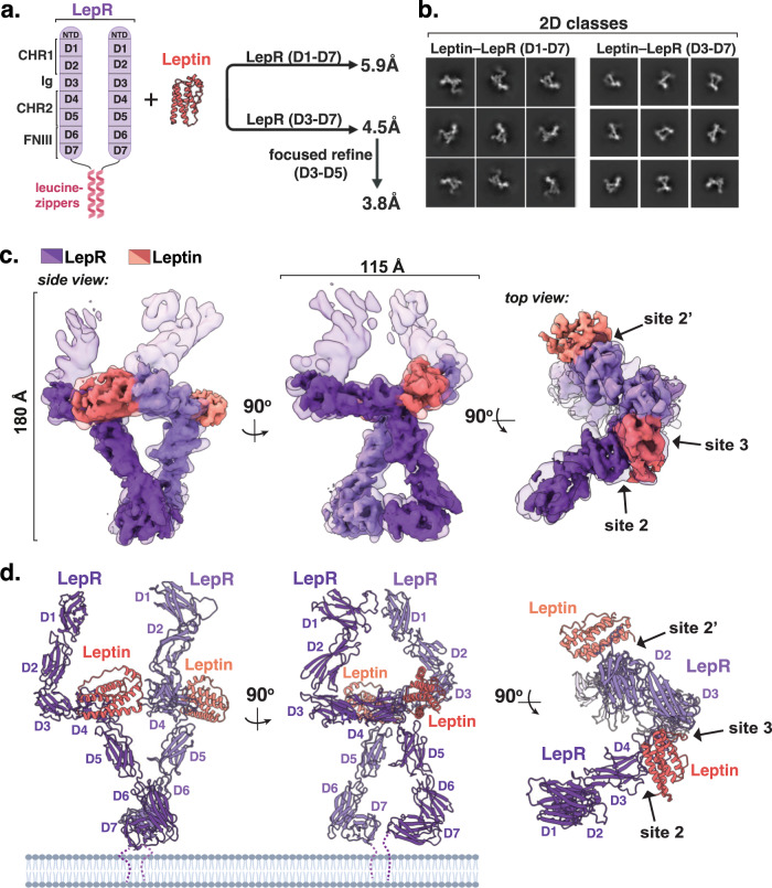
Leptin is an adipocyte-derived protein hormone that promotes satiety and energy homeostasis by activating the leptin receptor (LepR)-STAT3 signaling axis in a subset of hypothalamic neurons. Leptin signaling is dysregulated in obesity, however, where appetite remains elevated despite high levels of circulating leptin. To gain insight into the mechanism of leptin receptor activation, here we determine the structure of a stabilized leptin-bound LepR signaling complex using single particle cryo-EM. The structure reveals an asymmetric architecture in which a single leptin induces LepR dimerization via two distinct receptor-binding sites. Analysis of the leptin-LepR binding interfaces reveals the molecular basis for human obesity-associated mutations. Structure-based design of leptin variants that destabilize the asymmetric LepR dimer yield both partial and biased agonists that partially suppress STAT3 activation in the presence of wild-type leptin and decouple activation of STAT3 from LepR negative regulators. Together, these results reveal the structural basis for LepR activation and provide insights into the differential plasticity of signaling pathways downstream of LepR.


Conflict of interest statement

R.A.S and K.C.G are inventors on a patent application relating to engineered leptin variants described here. The remaining authors declare no competing interests.

Figures

Fig. 1
Fig. 1. Cryo-EM structure of the leptin receptor signaling complex.
a Schematic depicting LepR ECD domain architectures, and the regions analyzed for structure determination using cryo-EM. b Cryo-EM 2-dimensional class averages of assembled leptin-bound LepRD1–D7 and LepRD3–D7 complexes. c Overlaid segmented density maps of the Leptin-LepRD1–D7 and Leptin-LepRD3–D7 complexes resolved to 5.9 Å and 4.5 Å resolution, respectively. d Three views of the leptin-LepRD1–D7 structural model, with leptin in salmon and LepR in purple.
Fig. 2
Fig. 2. Structural homology between leptin and IL-6 family cytokine receptor complexes.
a Side and top views of leptin-bound LepR structural model (PDB ID: 8DH9), showing the leptin-binding domains (D3-D5) of LepR, with leptin in salmon and LepR in purple. b Side and top views of the hexameric IL-6 receptor complex (PDB ID: 1P9M) showing IL-6 in pink, gp130 in green, and IL-6Rα in gray. c Side and top views of the IL-27 receptor complex (PDB ID: 7U7N), showing IL-27 subunits p28 and Ebi3 in yellow and gray, respectively, gp130 in green, and IL-27Rα in blue. d Schematic showing how LepR mutants L503S/L504S (LepR-2KO) and L370S (LepR-3KO) assemble to exclusively form an asymmetric 1:2 leptin–LepR complex, in the same conformation as the partially open 2:2 complex observed in our structure. e Immunoblot of lysates prepared from HEK-293T cells transiently expressing the indicated LepR constructs and stimulated with 10 nM recombinant leptin for 20 min. Representative result of experiment performed three times.
Fig. 3
Fig. 3. Structural basis for leptin-dependent LepR dimerization.
a Front view of the segmented density map of the leptin-LepRD3-D7 complex resolved to 4.5 Å resolution (transparent) with the focus refined map encompassing leptin and the leptin-binding domains of LepR, resolved to 3.8 Å resolution (solid). b, c Close-up views of the leptin-LepR site 2 binding interface. Hydrogen bonds and salt-bridges are shown as black dashed-lines. d Immunoblot of lysates prepared from HEK-293T cells transiently expressing the indicated LepR constructs and stimulated with the indicated concentration of recombinant leptin for 20 min. Representative result of experiment performed two times. e Top view of the segmented density maps shown in (a). f Close-up view of the leptin-LepR site 3a binding interface. g Immunoblot of lysates prepared and analyzed as in (c). Representative result of experiment performed two times. h Comparison of the apo-leptin structure (PDB ID: 1AX8) and LepR-bound leptin structure (this paper, PDB ID: 8DHA), showing leptin in salmon, LepR D3 in purple. i Close-up view of the leptin-LepR site 3b binding interface. j LepR-dependent ordering of the leptin AB loop residues 24–39, shown in green. k Immunoblot of lysates prepared from HEK-293T cells stably expressing wild-type LepR and stimulated with the indicated leptin variants for 20 min. Representative result of experiment performed three times.
Fig. 4
Fig. 4. Biased leptin analogs decouple activation of STAT3 from LepR negative regulators.
a Cartoon model of LepR signaling in which STAT3 recruited by phosphorylation of LepR-Y1141, whereas LepR negative regulators are recruited by phosphorylation of LepR-Y986. b Phospho-STAT3 dose-response curves for WT or mutant leptin in LepR-expressing HEK 293T cells, analyzed by flow cytometry and shown as a percent of maximal WT leptin mean fluorescent intensity (MFI ± SEM, n = 4 (mutants) or 6 (WT) independent replicates). c Immunoblot of lysates prepared from HEK-293T cells stably expressing wild-type LepR that were serum starved for 18 h and then stimulated with the indicated leptin variants for 20 min. Representative result of experiment performed four times. d Quantification of immunoblots prepared as in (c) (100 nM leptin, mean ± SEM, n = 4 independent replicates, t-test, **p = 0.004, ***P < 0.001). e Schematic showing engineering strategy to create high affinity LepR partial agonists, through increasing affinity of leptin for LepR at site 2 and decreasing affinity at site 3. f Phospho-STAT3 signaling in LepR-expressing HEK 293T cells stimulated with 10 nM WT Leptin and the indicated concentration of leptin variants, analyzed as in (B) (mean ± SEM, n = 4 independent replicates). g Relative expression of SOCS3 from hLepR-expressing HEK 293T cells stimulated with 10 nM WT Leptin and 100 nM of the indicated leptin variants for 6 h, analyzed by RT-qPCR. (mean ± SEM, n = 6 independent replicates, t-test, **p = 0.003). h Immunoblot of lysates prepared from HEK-293T cells stably expressing wild-type hLepR that were serum starved for 18 h and then stimulated with 10 nM WT leptin and the indicated concentration of the indicated leptin variants for 20 min. Representative result of experiment performed four times. i Quantification of immunoblots prepared as in (h). Cells treated with 100 nM leptin variants (± SEM, n = 4 independent replicates, t-test, ***p < 0.001).

References

    1. Blüher M. Obesity: global epidemiology and pathogenesis. Nat. Rev. Endocrinol. 2019;15:288–298. doi: 10.1038/s41574-019-0176-8. - DOI - PubMed
    1. Hruby A, Hu FB. The epidemiology of obesity: a big picture. Pharmacoeconomics. 2015;33:673–689. doi: 10.1007/s40273-014-0243-x. - DOI - PMC - PubMed
    1. Müller TD, Blüher M, Tschöp MH, DiMarchi RD. Anti-obesity drug discovery: advances and challenges. Nat. Rev. Drug Discov. 2022;21:201–223. doi: 10.1038/s41573-021-00337-8. - DOI - PMC - PubMed
    1. Zhang Y, et al. Positional cloning of the mouse obese gene and its human homologue. Nature. 1994;372:425–432. doi: 10.1038/372425a0. - DOI - PubMed
    1. Friedman JM. Leptin and the endocrine control of energy balance. Nat. Metab. 2019;1:754–764. doi: 10.1038/s42255-019-0095-y. - DOI - PubMed

Publication types