Review
. 2023 Apr 13;66(7):4491-4502.
doi: 10.1021/acs.jmedchem.3c00072. Epub 2023 Apr 1.

Modulation of ERK5 Activity as a Therapeutic Anti-Cancer Strategy

Affiliations
Review

Modulation of ERK5 Activity as a Therapeutic Anti-Cancer Strategy

Duncan C Miller et al. J Med Chem. .

Abstract

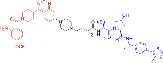
The extracellular signal-regulated kinase 5 (ERK5) signaling pathway is one of four conventional mitogen-activated protein (MAP) kinase pathways. Genetic perturbation of ERK5 has suggested that modulation of ERK5 activity may have therapeutic potential in cancer chemotherapy. This Miniperspective examines the evidence for ERK5 as a drug target in cancer, the structure of ERK5, and the evolution of structurally distinct chemotypes of ERK5 kinase domain inhibitors. The emerging complexities of ERK5 pharmacology are discussed, including the confounding phenomenon of paradoxical ERK5 activation by small-molecule ERK5 inhibitors. The impact of the recent development and biological evaluation of potent and selective bifunctional degraders of ERK5 and future opportunities in ERK modulation are also explored.


Conflict of interest statement

The authors declare no competing financial interest.

Figures

Figure 1
Figure 1
Schematic of the structure of ERK5 and the conformational changes that occur on phosphorylation, leading to nuclear translocation.
Figure 2
Figure 2
Structures of BIX02188 (1), BIX02189 (2), and TG02 (3).
Figure 3
Figure 3
(a) The structure of selected diazepinone ERK5 inhibitors. (b) X-ray cocrystal structure of a diazepinone inhibitor bound to ERK5 (PDB accession code 4B99). (c) Interaction map of 4 with ERK5. Key: (blue arrow) main chain donor/acceptor, (green circle) grease, (pink circle/blue outline/red outline) polar/basic/acidic, (circle blue eclipse) residue protection, (blue circle) solvent exposure.
Figure 4
Figure 4
Structures of selected pyrrole carboxamide ERK5 inhibitors.
Figure 5
Figure 5
(a) X-ray cocrystal structure of a pyrrole carboxamide inhibitor 11 bound to ERK5 (PDB accession code 7PUS). (b) Interaction map of pyrrole carboxamide inhibitor 11 with ERK5. Key: (blue arrow) main chain donor/acceptor, (green circle) grease, (pink circle/blue outline/red outline) polar/basic/acidic, (circle blue eclipse) residue protection, (blue circle) solvent exposure, (dashes) substitution contour.
Figure 6
Figure 6
(a) The structures of quinazoline-based ERK5 inhibitors 12 and 13 (BAY-885). (b) X-ray cocrystal structure of a quinazoline scaffold bound to ERK5 (PDB accession code 6HKN). (c) Interaction map of quinazoline inhibitor with ERK5. Key: (blue arrow) main chain donor/acceptor, (green arrow) side chain donor/acceptor, (gold arrow) water H bond, (white circle) water, (green circle) grease, (pink circle/blue outline/red outline) polar/basic/acidic, (circle blue eclipse) residue protection, (blue circle) solvent exposure, (dashes) substitution contour.
Figure 7
Figure 7
(a) The structures of thiophene dual-target inhibitors 14 and 15 (ADTL-EI1712). (b) Model of 14 bound to the active site of ERK5 using PDB accession code 4B99. Residues within 8 Å of 14 are colored using sequence conservation against ERK1 (green, conserved; red, nonconserved). (c) Potential interaction map of compound 14 bound with ERK5. Key: (blue arrow) main chain donor/acceptor, (green circle) grease, (pink circle/blue outline/red outline) polar/basic/acidic, (circle blue eclipse) residue protection, (blue circle) solvent exposure, (dashes) substitution contour.
Figure 8
Figure 8
Bivalent Inhibitor design. (a) Structure of 16 (ERK5.1). PP2-derived kinase ligand substructure shown in red. The D-site peptide motif is highlighted in blue. (b) Proposed binding mode of 16 bound to ERK5 engaging with the hinge and the D site.
Figure 9
Figure 9
Structure of ERK5 bifunctional degrader 17 (INY-06-061). Quinazoline-based ERK kinase inhibitor motif shown in red, linker shown in black, and VHL ligand shown in blue.

References

    1. Nithianandarajah-Jones G. N.; Wilm B.; Goldring C. E. P.; Müller J.; Cross M. J. ERK5: Structure, Regulation and Function. Cell. Signal. 2012, 24 (11), 2187–2196. 10.1016/j.cellsig.2012.07.007. - DOI - PubMed
    1. Lee J. D.; Ulevitch R. J.; Han J. H. Primary Structure of BMK1: A New Mammalian MAP Kinase. Biochem. Biophys. Res. Commun. 1995, 213 (2), 715–724. 10.1006/bbrc.1995.2189. - DOI - PubMed
    1. Zhou G.; Bao Z. Q.; Dixon J. E. Components of a New Human Protein Kinase Signal Transduction Pathway *. J. Biol. Chem. 1995, 270 (21), 12665–12669. 10.1074/jbc.270.21.12665. - DOI - PubMed
    1. Paudel R.; Fusi L.; Schmidt M. The MEK5/ERK5 Pathway in Health and Disease. Int. J. Mol. Sci. 2021, 22 (14), 7594.10.3390/ijms22147594. - DOI - PMC - PubMed
    1. Stecca B.; Rovida E. Impact of ERK5 on the Hallmarks of Cancer. Int. J. Mol. Sci. 2019, 20 (6), 1426.10.3390/ijms20061426. - DOI - PMC - PubMed

Publication types

MeSH terms

Substances

LinkOut - more resources