. 2023 Apr:90:104545.
doi: 10.1016/j.ebiom.2023.104545. Epub 2023 Mar 30.

Emergence and antibody evasion of BQ, BA.2.75 and SARS-CoV-2 recombinant sub-lineages in the face of maturing antibody breadth at the population level

Affiliations

Emergence and antibody evasion of BQ, BA.2.75 and SARS-CoV-2 recombinant sub-lineages in the face of maturing antibody breadth at the population level

Anouschka Akerman et al. EBioMedicine. 2023 Apr.

Abstract

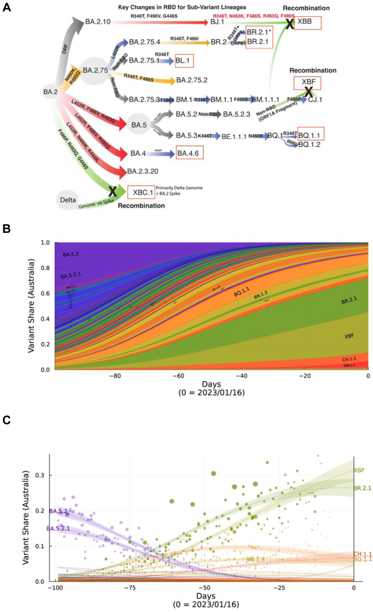
Background: The Omicron era of the COVID-19 pandemic commenced at the beginning of 2022 and whilst it started with primarily BA.1, it was latter dominated by BA.2 and the related sub-lineage BA.5. Following resolution of the global BA.5 wave, a diverse grouping of Omicron sub-lineages emerged derived from BA.2, BA.5 and recombinants thereof. Whilst emerging from distinct lineages, all shared similar changes in the Spike glycoprotein affording them an outgrowth advantage through evasion of neutralising antibodies.

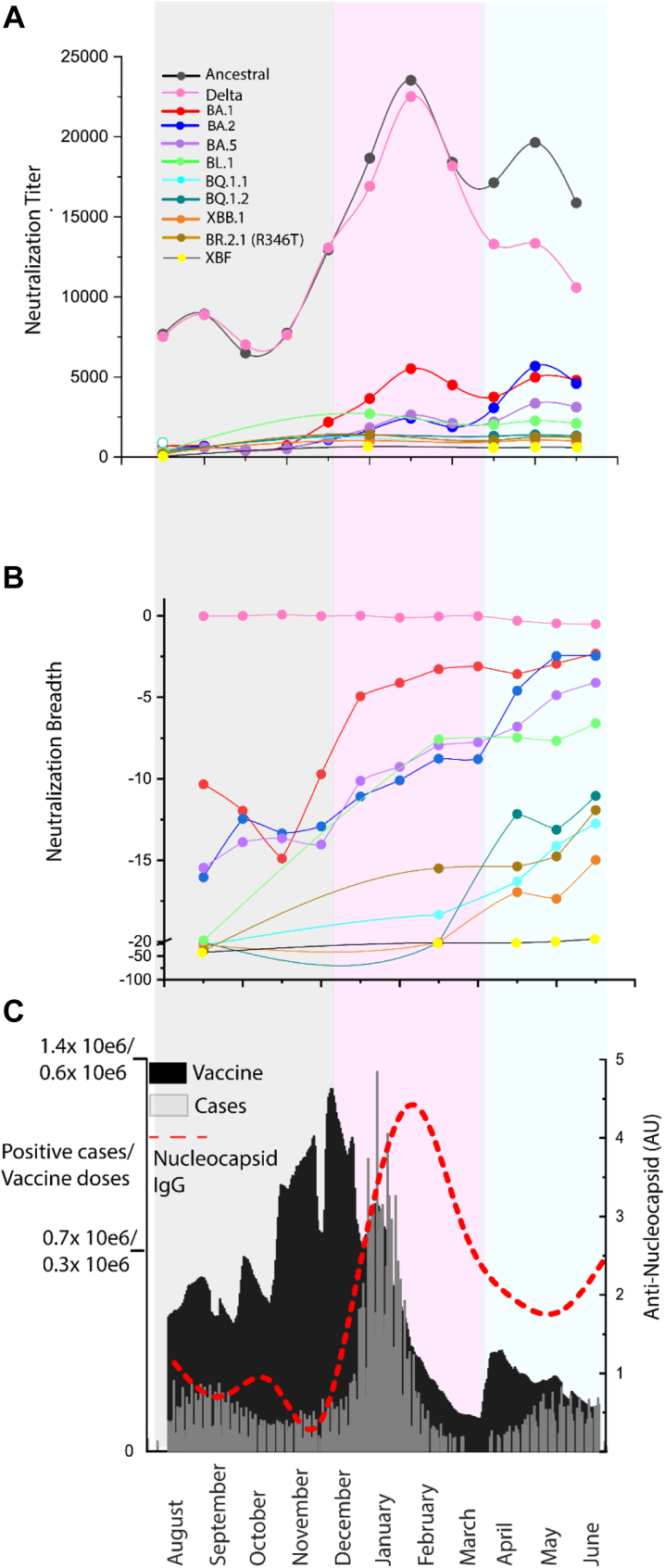
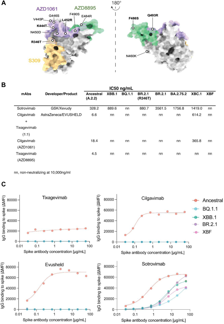
Methods: Over the course of 2022, we monitored the potency and breadth of antibody neutralization responses to many emerging variants in the Australian community at three levels: (i) we tracked over 420,000 U.S. plasma donors over time through various vaccine booster roll outs and Omicron waves using sequentially collected IgG pools; (ii) we mapped the antibody response in individuals using blood from stringently curated vaccine and convalescent cohorts. (iii) finally we determine the in vitro efficacy of clinically approved therapies Evusheld and Sotrovimab.

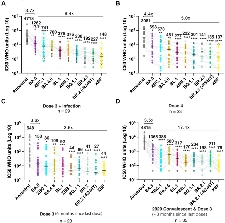
Findings: In pooled IgG samples, we observed the maturation of neutralization breadth to Omicron variants over time through continuing vaccine and infection waves. Importantly, in many cases, we observed increased antibody breadth to variants that were yet to be in circulation. Determination of viral neutralization at the cohort level supported equivalent coverage across prior and emerging variants with isolates BQ.1.1, XBB.1, BR.2.1 and XBF the most evasive. Further, these emerging variants were resistant to Evusheld, whilst increasing neutralization resistance to Sotrovimab was restricted to BQ.1.1 and XBF. We conclude at this current point in time that dominant variants can evade antibodies at levels equivalent to their most evasive lineage counterparts but sustain an entry phenotype that continues to promote an additional outgrowth advantage. In Australia, BR.2.1 and XBF share this phenotype and, in contrast to global variants, are uniquely dominant in this region in the later months of 2022.

Interpretation: Whilst the appearance of a diverse range of omicron lineages has led to primary or partial resistance to clinically approved monoclonal antibodies, the maturation of the antibody response across both cohorts and a large donor pools importantly observes increasing breadth in the antibody neutralisation responses over time with a trajectory that covers both current and known emerging variants.

Funding: This work was primarily supported by Australian Medical Foundation research grants MRF2005760 (SGT, GM & WDR), Medical Research Future Fund Antiviral Development Call grant (WDR), the New South Wales Health COVID-19 Research Grants Round 2 (SGT & FB) and the NSW Vaccine Infection and Immunology Collaborative (VIIM) (ALC). Variant modeling was supported by funding from SciLifeLab's Pandemic Laboratory Preparedness program to B.M. (VC-2022-0028) and by the European Union's Horizon 2020 research and innovation programme under grant agreement no. 101003653 (CoroNAb) to B.M.

Keywords: Covid-19; Evusheld; Neutralising antibodies; SARS-CoV-2; Sotrovimab; TMPRSS2; Variants.


Conflict of interest statement

Declaration of interests Funding bodies did not contribute to study design, data collection, data analysis or writing of the manuscript. Study design, data collection, data analysis and data interpretation was performed by the listed authors. Writing and review was performed by the listed authors. A.L.C. Participated on the AstraZeneca COVID-19 Advisory Board (2021), Seqirus COVID Advisory Board (2020) & CSL Seqirus APAC Advisory Council (2022). N.R., T.B., S.A.C., S.M., and T.H. are employees and shareholders of CSL LTD, a manufacturer of the polyclonal immunoglobulin preparations used as a reagent within the study.

Figures

Fig. 1
Fig. 1
Emergence and prevelance of BA.2.75 and BA.5 sub-lineages with convergent Spike polymorphisms in Australia. A. From left to right, Omicron lineages from the parent BA.2 are now primarily split across BA.2.75 and BA.5 sub-lineages. Divergent lineages such as BR.2.1 have accumulated polymorphisms common to BA.5, including L452R and F486I. Whilst BQ.1.1 has acquired BA.2.75 polymorphisms K444T and N460K. Common across most lineages is the additional acquisition of R346T, and this change defines lineages BL.1 and BA.4.6. XBB.1 is a recombinant between BJ.1 and BM.1.1.1 and has benefited from many Spike changes through this recombination event. Whilst XBF is a recombinant between BA.5.2.3 and CJ.1, it retains the latter Spike defined by the F486P change. XBC.1 is a Delta-BA.2 recombinant, with the retention of an intact BA.2 Spike with F486P, G446S and R493Q. Boxed in red are variants that were selected for testing in this study and range from variants with few spike polymorphisms (BL.1 and BA.4.6) through those with many convergent polymorphisms (BR.2.1, XBF and BQ.1.1). For the latter, we have also looked a two circulating isolates of BR.2.1, with and without the R346T change. B. Surveillance summary of Australia at 1601/2 based on genomic data via GISAID. Each vertical slice depicts the posterior mean variant frequency estimate from a hierarchical Bayesian multinomial logistic model of variant competition. Note the two dominant variants BR.2.1 and the recombinant XBF which are dominating in the states of NSW and Victoria, respectively. C. Variant proportions with associated Bayesian Credible Intervals for B.
Fig. 2
Fig. 2
Potency and breadth in pooled IgGs of greater than 420,000 U.S. plasma donors from August 2021 through to June 2022. A. Neutralization titers across 11 clinical isolates and include Clade A.2.2 (Ancestral), Delta and Omicron BA.1, BA.2, BA.5, BL.1, BQ.1.1, BQ.1.2, XBB.1, XBF and BR.2.1 (with R436T) for IgG batches collected between August 2021 and June 2022 (batch details are presented in Supplementary Table S1). Each point represents the mean titer of batches from that month and constitutes approximately 30,000 plasma donations. B. Breadth score of batches to variants in A. Breadth score is calculated as 1- (fold reduction of titer relative to the Clade A variant A.2.2). When the breadth approaches zero (e.g. Delta), neutralization titers cover this variant with equal potency to the ancestral variant. C. Documented vaccines doses and COVID-19 cases are presented here to frame the responses in A and B. Anti-nucleocapsid IgG levels in monthly batches presented in A and B are also indicated (red line). The grey-shading is primarily the 3× booster doses administered prior to the arrival of Omicron BA.1. In pink is the Omicron BA.1 wave. In blue is primarily the Omicron BA.2 wave.
Fig. 3
Fig. 3
Emerging variants and their ability to evade a continuum of antibody responses. A. Three dose Pfizer BNT162b2 vaccination with subsequent breakthrough infection. Closed circles were infections between June and August 2022 when BA.2 and BA.5 were prevalent. Open circles are breakthrough infections with BA.1 between January and February. B. Primarily four dose BNT162b2 vaccinations, in which the last dose was within three months. Open circles in this group are breakthrough infections in early 2022 at the time of BA.1. C. Three dose Pfizer BNT162b2 vaccination with the last dose six months prior. D. Early 2020 donors infected between March and August of 2020 and then subsequently vaccinated with two doses of Pfizer BNT162b2 or AstraZeneca AZD1222/ChAdOx1 and boosted with Pfizer BNT162b2 or Moderna mRNA-1273. This group did not receive their last dose three to six months prior. Data in (A–D) indicates the mean IC50 of technical replicates for individual samples. The median titers are labelled. Fold change reductions in IC50 neutralization titers compare variants of concern to the Ancestral variant and Omicron BA.5 where indicated. ∗p < 0.05, ∗∗p < 0.01, ∗∗∗p < 0.001, ∗∗∗∗p < 0.0001 for Friedman test with Dunn's multiple comparison test.
Fig. 4
Fig. 4
Neutralizing activity of monoclonal antibodies against emerging variants. A. Binding sites for AZD1061 (Cilgavimab), AZD8895 (Tixagevimab) and S309 (Sotrovimab) on the SARS-CoV-2 Spike glycoprotein. Key Spike polymorphisms that map to these sites are indicated and are prevelant on emerging variants tested. B. IC50 values (ng/μl) of monoclonal antibodies Sotrovimab, Cilgavimab and Tixagevimab cocktail, Cilgavimab alone and Tixagevimab alone, against ancestral A.2.2, Omicron XBB.1, BQ.1.1, BR.2.1 (R346T), BR.2.1, BA.2.75.2, XBC.1 and XBF. Antibodies used herein were clinical grade batches. Data represents mean IC50 values from two independent experiments. For Sotrovimab, pairwise comparisons between Ancestral and variants were done using Kruskal Wallis test with Dunn's multiple comparisons. For other datasets, unpaired t-test was used. p values are indicated in Supplementary Table S6. C. Antibody binding to full length Spike expressed on live cells and acquired using flow cytometry. Signal is expressed as Mean Fluorescent Intensity above background Fluorescence (ΔMFI).
Fig. 5
Fig. 5
Variant divergence in TMPRSS2 use in the emerging Omicron variant swarm. A. Endpoint titers for primary nasopharyngeal swabs grouped into variants derived from BQ (BQ1.X) lineages and variants derived from BA.2.75 lineages (primarily BR.2.1) using the HAT-24 cell line. The linear regressions of BA.2, BA.5 and Delta are also presented and serves here primarily as a guide with respect viral titers per diagnostic Ct value. Of note, as the variant uses TMPRSS2 more efficiently, there is an increase in viral titer per Ct value and this is observed by an upwards shift in the linear regression. B. Air liquid interface (ALI) primary bronchial and alveolar epithelial cells 3 and 6 days post-infection. Equal numbers of viral particles were used to inoculate each culture. Supernatant was harvested 3 and 6 days post-infection. Error bars represent standard deviations from the mean of infections from two to four independent donors. Delta and the parent variants BA.2 and BA.5 are presented here to representative highlight the influence of TMPRSS2 usage for viral entry fitness in primary differentiated respiratory epithelia in the upper and lower respiratory tract. ∗p < 0.01, ∗∗p < 0.01, ∗∗∗p < 0.001, for two-way ANOVA with Sidak's multiple comparisons C. BQ.1.2 and D. BR.2.1 primary nasopharyngeal swabs are presented in HAT-24 cells following 3 days in culture. Scale bar represents 100 μm. Limiting viral dilutions are presented to contrast the growth of each variant. E. To enable initial resolution of variants, we determined RNA copies per diagnostic Ct values and have presented infectivity to particle ratios for variants that were initially group in A. through dividing TCID50 titers with copies per reaction based on qPCR cycle threshold values and internal PCR controls for quantification. Grouping of BA.5 and BA.2.75 lineages here highlight the latter group to have lower infectivity to particle ratios. ∗∗p < 0.01 for Mann Whitney U-test. F. Mean fold reductions in viral titers in the presence of saturating levels of the TMPRSS2 inhibitor Nafamostat. Mean values represent three independent experiments and each point is the mean fold reduction per independent experiment performed in quadruplicate. ∗p < 0.05, ∗∗p < 0.01, ∗∗∗p < 0.001, for ordinary one way ANOVA with Dunnett's multiple comparison test.

References

    1. Tea F., Ospina Stella A., Aggarwal A., et al. SARS-CoV-2 neutralizing antibodies: longevity, breadth, and evasion by emerging viral variants. PLoS Med. 2021;18(7) - PMC - PubMed
    1. Karbiener M., Farcet M.R., Schwaiger J., et al. Plasma from post-COVID-19 and COVID-19-vaccinated donors results in highly potent SARS-CoV-2 neutralization by intravenous immunoglobulins. J Infect Dis. 2021;224(10):1707–1711. - PMC - PubMed
    1. Gaebler C., Wang Z., Lorenzi J.C.C., et al. Evolution of antibody immunity to SARS-CoV-2. Nature. 2021;591(7851):639–644. - PMC - PubMed
    1. Dan J.M., Mateus J., Kato Y., et al. Immunological memory to SARS-CoV-2 assessed for up to 8 months after infection. Science. 2021;371(6529) - PMC - PubMed
    1. Kreer C., Zehner M., Weber T., et al. Longitudinal isolation of potent near-germline SARS-CoV-2-neutralizing antibodies from COVID-19 patients. Cell. 2020;182(6):1663–1673. - PMC - PubMed

Supplementary concepts