. 2023 Apr 12;31(4):634-649.e8.
doi: 10.1016/j.chom.2023.03.007. Epub 2023 Mar 31.

Engineered Escherichia coli for the in situ secretion of therapeutic nanobodies in the gut

Affiliations

Engineered Escherichia coli for the in situ secretion of therapeutic nanobodies in the gut

Jason P Lynch et al. Cell Host Microbe. .

Abstract

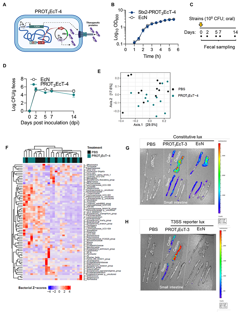
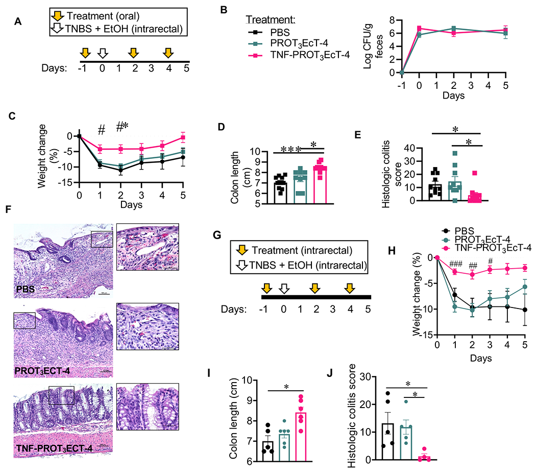
Drug platforms that enable the directed delivery of therapeutics to sites of diseases to maximize efficacy and limit off-target effects are needed. Here, we report the development of PROT3EcT, a suite of commensal Escherichia coli engineered to secrete proteins directly into their surroundings. These bacteria consist of three modular components: a modified bacterial protein secretion system, the associated regulatable transcriptional activator, and a secreted therapeutic payload. PROT3EcT secrete functional single-domain antibodies, nanobodies (Nbs), and stably colonize and maintain an active secretion system within the intestines of mice. Furthermore, a single prophylactic dose of a variant of PROT3EcT that secretes a tumor necrosis factor-alpha (TNF-α)-neutralizing Nb is sufficient to ablate pro-inflammatory TNF levels and prevent the development of injury and inflammation in a chemically induced model of colitis. This work lays the foundation for developing PROT3EcT as a platform for the treatment of gastrointestinal-based diseases.

Keywords: TNF; colitis; designer probiotic; engineered E. coli; in situ drug delivery; inflammatory bowel disease; live biotherapeutic; nanobody; type III secretion.


Conflict of interest statement

Declaration of interests C.F.L is on the scientific advisory board (SAB) for and A.Z.R. is an employee and has equity at Synlogic Therapeutics. J.P.L is an employee at AdAlta Ltd. W.S.G. is on the SABs of Kintai Therapeutics, SanaRx, Evelo Biosciences, and Tenza. F.I.S. is a co-founder and shareholder of Odyssey Therapeutics and a shareholder of IFM Therapeutics. C.B.S is a scientific advisor, and V.K. is the CEO and co-founder of Vicero, Inc. PROT(3)EcT is the subject of US Patent no. 9,951,340 and US Patent 10,702,559, both filed by Massachusetts General Hospital.

Figures

Figure 1.
Figure 1.. E. coli engineered with a modified T3SA efficiently secrete proteins into their surroundings.
(A, B, C, E, F) Secretion assays of designated strains engineered to secrete OspC2-FLAG. Supernatant (TCA precipitated) (S) and whole-cell pellet lysate (P) fractions were obtained 30-minutes (A, B, E), 6-hours (C), or at times indicated (F) post-transfer to new media. Unless otherwise indicated in (A), IPTG was present to induce expression of VirB and OspC2. Congo red (CR) was added, when noted. Immunoblots labeled with anti-FLAG or anti-GroEL antibodies (A, B, E, F) or a Coomassie blue-stained gel (C) are shown. (D) Schematic of PROT3EcT-1 that expresses plasmid-encoded Ptrc VirB and Ptrc OspC2-FLAG. (G) Gentamicin protection assay to assess invasion of bacteria into intestinal epithelial cells (HCT8). Each dot represents a biological replicate, and the horizontal bar indicates the mean. Data were analyzed using one-way ANOVA with Tukey’s post-hoc test; for all strain comparisons to Shigella *** P < 0.0001. (H) Translocation assay to assess the injection of OspC2-FLAG into cervical epithelial cells (HeLa). The soluble and insoluble (bacteria-containing) fractions were separated and immunoblotted with anti-FLAG and anti-ß-actin. Data in each panel are representative of at least 2 independent experiments. See also Figure S1.
Figure 2.
Figure 2.. PROT3EcT can be engineered to secrete nanobodies.
Secretion assays of (A) NbASC fused to designated N-terminal type III secretion signals and a C-terminal HA-tag, (B) NbASC, -NbPD-L1, -NbCTLA-4 and -NbNP1 fused to the N-terminal OspC2 secretion signal (SSOspC2) and C-terminal HA-tag, (C) monomeric (1x), heterodimeric (2x) and heterotrimeric (3x) NbStx2, fused to an N-terminal SSOspC2and a C-terminal 3xFLAG tag, (D) NbStx2 dimer modified with an N-terminal SSOspC2 (SSOspC2-Nb2x) and C-terminal HA tag. (A, B, C) were obtained at 6-hours. Immunoblots labeled with anti-GroEL as well as anti-HA (A, B) or anti-FLAG (C) antibodies or (D) a Coomassie blue-stained gel are shown. (E) Stx2 ELISA. (F) Stx2 Vero killing assay. For (E-F), each dot represents an individual biological replicate. Data in each panel are representative of at least 2 independent experiments. See also Figure S1.
Figure 3.
Figure 3.. PROT3EcT stably colonizes the gastrointestinal tract of mice but does not affect the gut microbiota.
(A) Schematic of PROT3EcT-4, which expresses VirB via a constitutive promoter (Pc) from a chromosomal locus. The Nb is expressed via a constitutive promoter from a plasmid maintained via auxotrophic selection. (B) Growth curves of wild-type EcN and Stx2-PROT3EcT grown in parallel. Data are presented as the mean ± SEM. (C) Study design schematic. (D) Shed bacterial titers. Data are presented as the mean ± SEM, each with 4 mice per group, and reflect at least 2 independent experiments. (E) Principal Coordinate Analysis (PCoA) plot showing Bray-Curtis dissimilarity for 16S rRNA gene amplicon fecal surveys from sham-treated (PBS) (n=12) and PROT3EcT-treated (n=12) mice, collected on day 14 post-treatment. (F) A hierarchically clustered heatmap of the z-score abundance of bacterial family/genus (f = family, g = genus) within each sample (rows) from the same samples as in (E). Color codes indicate treatment conditions (PBS vs. PROT3EcT). (G-H) Bioluminescent imaging of intestinal explants isolated from individual mice 8 dpi inoculated orally with designated strains transformed with (D) a constitutive bioluminescent reporter (pMM543) or (E) a T3SS-dependent reporter system (pMxiE-lux+ and pNG162-IpgC). Starting 1 day before they were administered bacteria to ensure plasmid maintenance, the mice began receiving kanamycin and spectinomycin in their drinking water. See also Figures S1–S2 and Tables S1A–B.
Figure 4.
Figure 4.. TNF-PROT3EcT secretes fully functional anti-TNF Nbs and colonizes mice but does not affect the gut microbiota.
(A) Secretion assays of PROT3EcT-1 engineered with monomeric (1x) or homodimeric (2x) NbTNF fused to an N-terminal OspC2ss and a C-terminal FLAG. Supernatant (TCA precipitated) (S) and whole-cell pellet lysate (P) fractions were obtained at 6-hours. Immunoblots labeled with an anti-FLAG antibody are shown. (B) Viability of L929 cells following incubation with 0.2 ng/ml of murine TNFα plus either supernatant (Sup) from designated strains or purified homodimeric NbTNF. Data are presented as the mean ± SEM (C) Shed bacterial titers. Each dot represents an individual mouse, and the horizontal bar indicates the mean. (D) PCoA plot showing Bray-Curtis dissimilarity for 16S rRNA gene amplicon fecal surveys from PROT3EcT (n=7) and TNF-PROT3EcT-treated (n=8) mice. (E) A hierarchically clustered heatmap of the z-score abundance of bacterial family/genus (f = family, g = genus) within each sample (rows) from the same samples as in (D). Color codes indicate treatment group (PROT3EcT vs. TNF-PROT3EcT) and caging information. Data in each panel are representative of at least 2 independent experiments. See also Figures S3–S5 and Tables S2 and S3A–B.
Figure 5.
Figure 5.. TNF-PROT3EcT inhibits the development of TNBS-induced colitis.
(A, G) Study design schematic. (B) Shed bacterial titers. (C, H) Body weight change (%). In (C), *denotes comparison to PBS group, P=0.0118; # denotes comparison to PROT3EcT-4; day 1, P=0.0238; day 2, P=0.0122. In (H), # denotes comparison to PROT3EcT-4, day 1, P=0.0003, day 2, P=0.0018, day 3, P=0.02. (D, I) Colon length. In (D), *, P=0.0219; ***, P=0.0004. In (I), *, P=0.0123. (E, J) Histologic colitis scores. In (E), vs. PBS *, P=0.0231; vs. PROT3EcT-4 *, P=0.0141. In (J), vs. PBS, P= 0.0406 and vs. PROT3EcT-4, P=0.0331. (F) Representative colon histology sections stained with hematoxylin and eosin. Scale bars = 100 μm. Data reflect at least 2 independent experiments, each with 3-5 mice per group, and are presented as mean ± SEM (B-C, H) or individual values ± SEM (D-E, I-J). Data were analyzed with a two-way ANOVA with Tukey’s post hoc test (B-C, H) or a Kruskal-Wallis test with Dunn’s multiple correction test (D-E, I-J). TNBS = 2,4,6-Trinitrobenzenesulfonic acid. EtOH = ethanol. See also Figures S4 and S6–7.
Figure 6.
Figure 6.. A single prophylactic dose of TNF-PROT3EcT attenuates TNBS-induced colitis.
(A) Study design schematic. (B) Shed bacteria titers. (C) Body weight change (%). *denotes comparison to PBS, day 1, P=0.0002, day 2, P<0.0001; #denotes comparison to PROT3EcT-4, day 1, P=0.054, day 2, P<0.0001. (D) Colon length. *, P=0.0184; **, P=0.0029. (E) Histologic colitis scores. **, P=0.0045. (F-H) Colon homogenates were analyzed for the levels of TNFα (F) (***, P=0.0005, *, P=0.0433), IL-6 (G) (vs. PROT3EcT-4 *, P=0.0356; vs. PBS *, P=0.0322) and IL-10 (H) by ELISA. Data reflect 2 independent experiments, each with 4-5 mice per group, and are presented as individual values ± SEM (D-G) or mean ± SEM (B-C). Data were analyzed using a two-way ANOVA with Tukey’s post hoc test (B-C) or a Kruskal-Wallis test with Dunn’s multiple correction test (D-H). TNBS = 2,4,6-Trinitrobenzenesulfonic acid. EtOH = ethanol. See also Figure S6.

References

    1. Basso PJ, Câmara NOS, and Sales-Campos H (2019). Microbial-Based Therapies in the Treatment of Inflammatory Bowel Disease – An Overview of Human Studies. Frontiers in Pharmacology 9. 10.3389/fphar.2018.01571. - DOI - PMC - PubMed
    1. Durack J, and Lynch SV (2019). The gut microbiome: Relationships with disease and opportunities for therapy. J Exp Med 216, 20–40. 10.1084/jem.20180448. - DOI - PMC - PubMed
    1. Cubillos-Ruiz A, Guo T, Sokolovska A, Miller PF, Collins JJ, Lu TK, and Lora JM (2021). Engineering living therapeutics with synthetic biology. Nat Rev Drug Discov 20, 941–960. 10.1038/S41573-021-00285-3. - DOI - PubMed
    1. McNerney MP, Doiron KE, Ng TL, Chang TZ, and Silver PA (2021). Theranostic cells: emerging clinical applications of synthetic biology. Nature Reviews Genetics 22, 730–746. 10.1038/s41576-021-00383-3. - DOI - PMC - PubMed
    1. Riglar DT, and Silver PA (2018). Engineering bacteria for diagnostic and therapeutic applications. Nat Rev Microbiol 16, 214–225. 10.1038/nrmicro.2017.172. - DOI - PubMed

Substances