. 2023 May 15;435(10):168070.
doi: 10.1016/j.jmb.2023.168070. Epub 2023 Mar 31.

Context-dependence of T-loop Mediated Long-range RNA Tertiary Interactions

Affiliations

Context-dependence of T-loop Mediated Long-range RNA Tertiary Interactions

Lisa N Hansen et al. J Mol Biol. .

Abstract

The architecture and folding of complex RNAs is governed by a limited set of highly recurrent structural motifs that form long-range tertiary interactions. One of these motifs is the T-loop, which was first identified in tRNA but is broadly distributed across biological RNAs. While the T-loop has been examined in detail in different biological contexts, the various receptors that it interacts with are not as well defined. In this study, we use a cell-based genetic screen in concert with bioinformatic analysis to examine three different, but related, T-loop receptor motifs found in the flavin mononucleotide (FMN) and cobalamin (Cbl) riboswitches. As a host for different T-loop receptors, we employed the env8 class-II Cbl riboswitch, an RNA that uses two T-loop motifs for both folding and supporting the ligand binding pocket. A set of libraries was created in which select nucleotides that participate in the T-loop/T-loop receptor (TL/TLR) interaction were fully randomized. Library members were screened for their ability to support Cbl-dependent expression of a reporter gene. While T-loops appear to be variable in sequence, we find that the functional sequence space is more restricted in the Cbl riboswitch, suggesting that TL/TLR interactions are context dependent. Our data reveal clear sequence signatures for the different types of receptor motifs that align with phylogenic analysis of these motifs in the FMN and Cbl riboswitches. Finally, our data suggest the functional contribution of various nucleobase-mediated long-range interactions within the riboswitch subclass of TL/TLR interactions that are distinct from those found in other RNAs.

Keywords: RNA structure; T-loop; regulatory RNA; structural motif; tertiary interaction.


Conflict of interest statement

Declaration of Interest

R.T.B. serves on the Scientific Advisory Boards of Expansion Therapeutics, SomaLogic and MeiraGTx.

Figures

Figure 1.
Figure 1.. Structure of the FMN/Cbl TL/TLR interaction.
(A) Structure of the classic T-loop (TL) motif showing the nucleobase configuration of the five nucleotides (positions 1–5). Panel on the left represents a front view emphasizing all nucleobases and the right is a 90° clockwise rotation of the left view emphasizing the stacking interactions and the space between positions 4 and 5. (B) Schematic of the terminal loop in FMN/Cbl riboswitches harboring the T-loop motif (red) and two bulged nucleotides (magenta). (C) Cartoon representation of the global architecture of the FMN riboswitch bound to FMN (green) (PDB 3F2Q). The T-loop motifs are shown in red, the two nucleotides 3′ of the TL in the terminal loop are magenta, the T-loop receptor is colored cyan, and the two nucleotides 3′ of the TLR in the terminal loop are dark blue along with two nucleotides immediately 3′ of the helix of the T-loop stem-loop that form a two-nucleotide helix (auxiliary helix). This color scheme will be used throughout this work. (D) Cartoon representation of the env8 Cbl riboswitch bound to Cbl (green). The TL/5nt-TLR interaction is shown in the same colors as in panel C, with a second T-loop motif and two interacting nucleotides that support the ligand binding pocket are shown in yellow and orange, respectively (PDB 4FRN). (E) Closeup view of the TL/5nt-TLR interaction in the env8 aptamer (PDB 4FRG). Color scheme is the same as in panels C and D. The four nucleobase-mediated contacts facilitated by the T-loop and the 5nt-TLR are labeled as i-iv.
Figure 2.
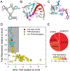
Figure 2.. Genetic screen of the env8 T-loop and associated nucleotides.
(A) Secondary structure of env8 aptamer domain with nucleotide positions represented as open circles and the fully randomized regions in the library are colored as in Figure 1. (B) Cartoon representation closeup of the TL/5nt-TLR interaction emphasizing the architecture of the randomized nucleotides (PDB 4FRG). (C) Same perspective as in panel B but showing the nucleobase-mediated hydrogen bonding network between the T-loop and the 5nt-TLR for the nucleotides randomized in this library. (D) Plot of the performance of the sequence variants that emerged from the screen. Individual variants are colored red (env8, considered as wild type against which all other variants are normalized), sequence variants found in the sequences of TL/5nt-TLRs in the FMN riboswitch family are cyan, and sequence variants for those not observed in FMN sequence space are colored yellow. Gray box marks those variants with strong performance (i.e., relative fluorescent units (RFU) <3.0 relative to env8 and Fold Repression >0.5 relative to env8). (E). Pie chart of the core five nucleotide T-loops found in the FMN family that interact with 5nt-TLRs. Each slice represents the fraction of the population that comprises a specific sequence and the color of each slice represents the population-weighted average of the fold repression activity normalized to env8 for observed variants that have that specific T-loop sequence. Gray slices represent sequence space not observed in the selection. Curves around pie chart denote overarching sequence motifs. Green and cyan marks denote TGNRA and CGNRA motifs, respectively.
Figure 3.
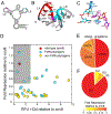
Figure 3.. Genetic screen of the 5nt-TLR.
(A) Secondary structure of env8 aptamer domain with nucleotide positions represented as open circles and the fully randomized regions in the library are colored as in Figure 1. (B) Cartoon representation closeup of the Cbl TL/5nt-TLR interaction emphasizing the architecture of the randomized nucleotides (PDB 4FRG). The numbering of nucleotides is consistent with that of the L4-L6 interaction in the env8 riboswitch. (C) Stick representation of the nucleobase-mediated hydrogen bonding network between the T-loop and the 5nt-TLR for the nucleotides randomized in this library. (D) Activity plot of the variants observed in the genetic screen. (E) Pie chart of the population distribution of the core five nucleotide sequence motif of the FMN 5nt-TLR. In panels D and E, the color scheme is the same as in Figure 2.
Figure 4.
Figure 4.. Genetic screen of the 4nt-TLR.
(A) Secondary structure of env8 aptamer domain with nucleotide positions represented as open circles and the fully randomized regions in the library are colored as in Figure 1. The circle with the dashed line represents the nucleotide deleted in the library. (B) Cartoon representation closeup of the TL-4ntTLR interaction emphasizing the architecture of the randomized nucleotides. (PDB 3F4E) The numbering of nucleotides is consistent with that of the L2-L6 interaction in the FMN riboswitch. (C) Stick representation of the nucleobase-mediated hydrogen bonding network between the T-loop and the 4nt-TLR for the nucleotides randomized in this library. (D) Activity plot of the variants observed in the genetic screen. (E) Pie chart of the population distribution of the core five nucleotide sequence motif of the FMN 4nt-TLR. (F) Pie chart of two nucleotide sequence motif of the FMN T-loop that interacts with the FMN 4nt-TLR. In panels D-F, the color scheme is the same as in Figure 2.
Figure 5.
Figure 5.. Genetic screen of the IL-TLR.
(A) Secondary structure of env8 aptamer domain with nucleotide positions represented as open circles and the fully randomized regions in the library are colored as in Figure 1. Note that the secondary structure has changed to reflect the conversion of the L6 terminal loop into an internal loop motif. (B) Cartoon representation closeup of the TL/IL-TLR interaction emphasizing the architecture of the randomized nucleotides. (PDB 4GXY) (C) Cartoon representation closeup of TL/IL-TLR interaction emphasizes randomized area. (D) Activity plot of the variants observed in the genetic screen. (E) Pie chart of the population distribution of the core five nucleotide sequence motif of the FMN IL-TLR. (F) Pie chart of population distribution of FMN riboswitches with the consensus URA/RA (left) and URU/RA (right) with associated activity. (G) Pie chart of the population distribution of sequences of J7/6 for those FMN IL-TLR switches with UAA J6/7 with associated activity. In panels D-G, the color scheme is the same as in Figure 2.
Figure 6.
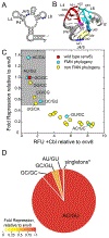
Figure 6.. Genetic screen of the auxiliary helix.
(A) Secondary structure of env8 aptamer domain with nucleotide positions represented as open circles and the fully randomized regions in the library are colored as in Figure 1. (B) Cartoon representation closeup of the TL/5nt-TLR interaction emphasizing the architecture of the randomized nucleotides. (PDB 4FRG) (C) Activity plot of the variants observed in the genetic screen. (D) Pie chart of the population distribution of the auxiliary helix from the FMN TL/5nt-TLR alignment. In panels C and F, the color scheme is the same as in Figure 2.
Figure 7.
Figure 7.. Assay of the common TL/5nt-TLR sequences in the FMN riboswitches.
Analysis of the most common sequences in the FMN riboswitch family yielded a set (v1-v11; inset) that comprises ~30% of all sequences. Nucleotide positions that differ from the wild type env8 sequence in the terminal loops are highlighted in green, except positions 2 and 3 in the auxiliary helix which are always a C-G base pair rather than the G-C pair in env8. The performance of each variant is plotted as previously, and the labels of the plot correspond to the sequence labels in the inset.

Comment in

References

    1. Hendrix DK, Brenner SE, Holbrook SR. RNA structural motifs: building blocks of a modular biomolecule. Q Rev Biophys. 2005;38:221–43. - PubMed
    1. Leontis NB, Lescoute A, Westhof E. The building blocks and motifs of RNA architecture. Curr Opin Struct Biol. 2006;16:279–87. - PMC - PubMed
    1. Holbrook SR. Structural principles from large RNAs. Annu Rev Biophys. 2008;37:445–64. - PubMed
    1. Butcher SE, Pyle AM. The molecular interactions that stabilize RNA tertiary structure: RNA motifs, patterns, and networks. Acc Chem Res. 2011;44:1302–11. - PubMed
    1. Batey RT, Rambo RP, Doudna JA. Tertiary Motifs in RNA Structure and Folding. Angew Chem Int Ed Engl. 1999;38:2326–43. - PubMed

Publication types