. 2023 Mar 31;11(1):66.
doi: 10.1186/s40168-023-01508-y.

Diet prevents the expansion of segmented filamentous bacteria and ileo-colonic inflammation in a model of Crohn's disease

Affiliations

Diet prevents the expansion of segmented filamentous bacteria and ileo-colonic inflammation in a model of Crohn's disease

Amira Metwaly et al. Microbiome. .

Abstract

Background: Crohn's disease (CD) is associated with changes in the microbiota, and murine models of CD-like ileo-colonic inflammation depend on the presence of microbial triggers. Increased abundance of unknown Clostridiales and the microscopic detection of filamentous structures close to the epithelium of Tnf ΔARE mice, a mouse model of CD-like ileitis pointed towards segmented filamentous bacteria (SFB), a commensal mucosal adherent bacterium involved in ileal inflammation.

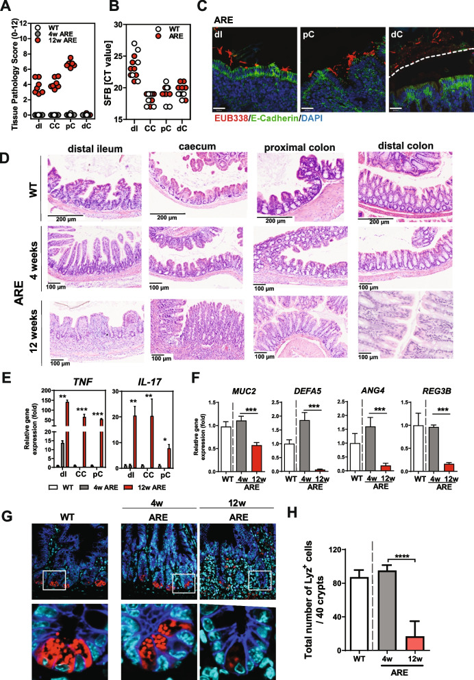
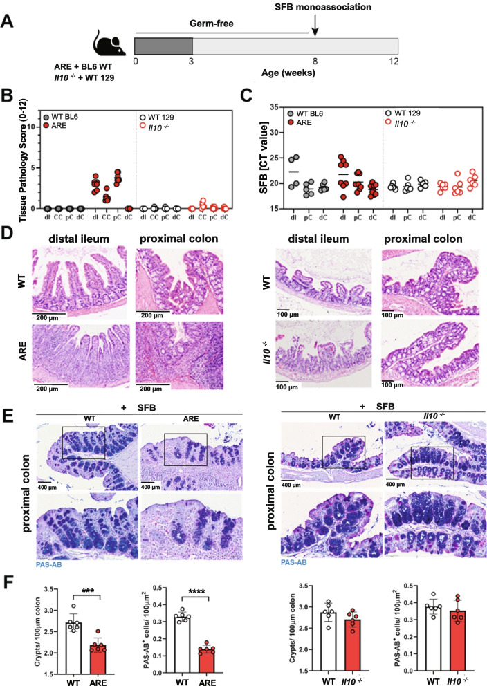
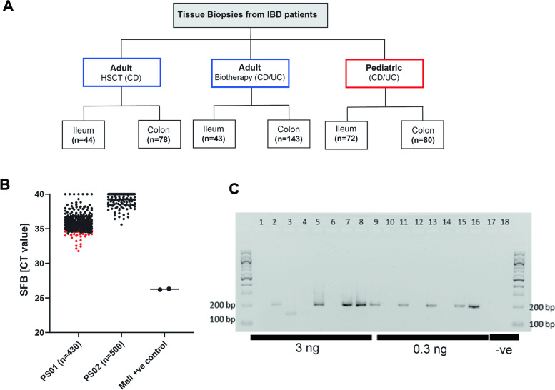
Results: We show that the abundance of SFB strongly correlates with the severity of CD-like ileal inflammation in two mouse models of ileal inflammation, including Tnf ΔARE and SAMP/Yit mice. SFB mono-colonization of germ-free Tnf ΔARE mice confirmed the causal link and resulted in severe ileo-colonic inflammation, characterized by elevated tissue levels of Tnf and Il-17A, neutrophil infiltration and loss of Paneth and goblet cell function. Co-colonization of SFB in human-microbiota associated Tnf ΔARE mice confirmed that SFB presence is indispensable for disease development. Screening of 468 ileal and colonic mucosal biopsies from adult and pediatric IBD patients, using previously published and newly designed human SFB-specific primer sets, showed no presence of SFB in human tissue samples, suggesting a species-specific functionality of the pathobiont. Simulating the human relevant therapeutic effect of exclusive enteral nutrition (EEN), EEN-like purified diet antagonized SFB colonization and prevented disease development in Tnf ΔARE mice, providing functional evidence for the protective mechanism of diet in modulating microbiota-dependent inflammation in IBD.

Conclusions: We identified a novel pathogenic role of SFB in driving severe CD-like ileo-colonic inflammation characterized by loss of Paneth and goblet cell functions in Tnf ΔARE mice. A purified diet antagonized SFB colonization and prevented disease development in Tnf ΔARE mice in contrast to a fiber-containing chow diet, clearly demonstrating the important role of diet in modulating a novel IBD-relevant pathobiont and supporting a direct link between diet and microbial communities in mediating protective functions. Video Abstract.

Keywords: Crohn’s disease; IBD; Inflammation; Pathobiont; Purified diet; Segmented filamentous bacteria; Tnf ΔARE mice.


Conflict of interest statement

The authors declare that they have no competing interests.

Figures

Fig. 1
Fig. 1
SFB abundance correlates with ileitis severity in SPF-house Tnf ΔARE mice. A Ileitis scores of SPF-housed Tnf ΔARE mice at the age of 3–4, 8, 12, and 18 weeks of age. Color-code represents severity of inflammation as described above. Cecal content of SPF-housed WT mice with undetectable SFB counts was introduced into germ-free Tnf ΔARE mice and littermate WT controls. B Quantitative analysis of SFB abundance in Tnf ΔARE and matching WT mice overtime. CT value > 30 is regarded as non-specificity threshold. C Representative H&E-stained tissue sections from distal ileum, caecum, and proximal colon of Tnf ΔARE mice showing no (NR), low (LR), and high grade (R) of inflammation in the ileum as described above. D Analysis of bacterial localization in distal ileum of inflamed (R) and non-inflamed (NR) Tnf ΔARE mice from F1 generation by performing FISH staining with eubacterial 16S probe (red) and DAPI for nuclear visualization. E Quantitative analysis of Tnf and Il-17A mRNA expression in distal ileum of non-inflamed (NR) and inflamed (R Tnf ΔARE mice and WT controls. F Correlation analysis between Ileitis score and SFB abundance in intestinal contents and fecal pellets in the full cohort of 18-week-old Tnf ΔARE mice and G in 12 10-week-old (mild ileitis) and 12 > 24-week-old (severe ileitis) SAMP1/YitFc mice, respectively. We replaced all data points below the detection limit (threshold of Ct > 30) by a fixed minimal value Ct = 30 in (F, G). This illustrates the dataset more clearly than excluding the values under the detection limit completely from the analysis. Statistical significance was assessed by One-way-ANOVA followed by Tukey multiple comparison test. *p < 0.05; **p < 0.01; ***p < 0.001 was considered statistically significant
Fig. 2
Fig. 2
SFB mono-colonization causes CD-like inflammation in Tnf ΔARE mice. Germ-free Tnf ΔARE mice and littermate WT controls were monocolonized with SFB from birth. A Severity of inflammation was assessed by evaluating the tissue pathology score in tissue sections of distal ileum, cecum, and proximal and distal colon from 12-week-old WT mice as well as 4-week-old and 12-week-old Tnf ΔARE mice. B Quantitative analysis of SFB abundance in intestinal contents from distal ileum, cecum, and proximal and distal colon of 12-week-old WT and Tnf ΔARE mice. C FISH by using the eubacterial probe EUB338 was performed on tissue sections from distal ileum and colon of 12-week-old Tnf ΔARE mice followed by immunostaining with E-Cadherin. DAPI was used for nuclear visualization. Dotted line indicates thickness of inner mucus layer. D H&E-stained tissue sections of 12-week-old Tnf ΔARE mice revealed profound intestinal inflammation in the compartments of distal ileum, cecum, proximal colon, but not jejunum and distal colon when compared to 12-week-old WT controls, but also 4-week-old Tnf ΔARE mice. Scale bar represents 200 or 100 μm as indicated. E Quantitative analysis of Tnf and Il-17A transcript levels in mucosal tissue of distal ileum, cecum, and proximal colon from 4 and 12-week-old Tnf ΔARE mice and 12-week-old WT controls. Statistical significance was assessed by One-way analysis of variance (ANOVA) followed by Tukey test. *p < 0.05; **p < 0.01; ***p < 0.001 was considered statistically significant. F Quantitative analysis of Muc2, Defa5, Ang4, and Reg3b transcript levels in mucosal tissue of distal ileum from 4- and 12-week-old Tnf ΔARE mice and 12-week-old WT controls. Statistical significance was assessed by One-way analysis of variance (ANOVA) followed by Tukey test. *p < 0.05; **p < 0.01; ***p < 0.001 was considered statistically significant. G Immunofluorescence (IF) co-staining of Lysozyme (red) and E-cadherin (IEC borders, blue) counterstained with Dapi (nuclei, cyan) in ileal tissue sections from 4 weeks and 12 weeks mono-colonized WT and Tnf ΔARE mice (× 600) lower panel: higher magnification of the indicated sections (× 3600). H Quantification of the total number of Lysozyme positive Paneth cells per crypt based on IF staining. Statistical analyses were performed by one-way analysis of variance (ANOVA) followed by Tukey test
Fig. 3
Fig. 3
SFB fail to induce ileo-colonic inflammation in a colitis mouse model. A GF Tnf ΔARE, Il10−/− mice and WT controls were mono-colonized with SFB from 8 to 12 weeks of age. B Severity of inflammation was assessed by evaluating the tissue pathology score in tissue sections of cecum, proximal, and distal colon from Tnf ΔARE mice and Il10−/− mice, respectively. C Quantitative analysis of SFB abundance in intestinal contents collected from ileal, colonic, and cecal compartments of 12-weeks-old Tnf ΔARE mice and Il10−/− mice, respectively. D H&E-stained tissue sections of Tnf ΔARE mice revealed intestinal inflammation in the compartments of distal ileum, cecum, proximal colon, compared to Il10−/− mice. Scale bar represents 200 or 100 μm as indicated. E, F PAS-AB goblet cell staining in proximal colon tissue sections from mono-colonized Tnf ΔARE and Il10−/−mice (× 400). Lower panel represents higher magnification (× 1200) of the indicated sections. Graph represents the quantification of goblet cells per crypt. Statistical analyses were performed by unpaired Student’s t test
Fig. 4
Fig. 4
PCR detection of segmented filamentous bacteria in tissue biopsies of patients with Inflammatory bowel diseases. A Human cohorts screened for the presence of SFB.The analyzed samples included mucosal biopsies collected from three IBD patient cohorts enrolled in Barcelona, Paris and Munich. The Spanish (HSCT) cohort included patients enrolled in follow-up for CD following treatment with autologous hematopoietic stem cell transplantation (AHSCT). Ileal (n=44) and colonic biopsies (n=86) from CD patients were included in the analysis. The French (Biotherapy) cohort included longitudinally collected samples from CD and UC patients who are treated with anti-TNF and other biotherapy drugs and recruited in Paris. Ileal (n=43) and colonic biopsies (n=143) from CD and UC patients were included in the analysis. The German pediatric cohort is a longitudinal prospective study comprising children with IBD and were recruited in Munich. Ileal (n=72) and colonic biopsies (n=80) were included in the analysis. A positive control of DNA extracted from a sample positive for SFB positive based on 16S sequencing (Mali, provided by Pamela Schnupf) B Quantitative analysis of SFB abundance in tissue biopsies using two primer sets (published by Jonsson [29] and by Snel [54] and the newly designed primer set; unpublished data, Pamela Schnupf). Quantification of SFB in human tissue biopsies was performed using the MasterMix SensiFAST™ SYBR (BIOLINE) and 3 different primer sets: for_5‘-TGTGGGTTGTGAATAGCAAT-3‘, rev_5‘-GCGAGCTTCCCTCATTACAAGG-3’, for_5‘-AGGAGGAGTCTGCGGCACATTAGC-3‘ ,rev_5‘ TCCCCACTGCTGCCTCCCGTAG-3‘  and for,_ 5’-TGTAGGTTGTGAAGAACAAT-3’, rev_ 5’-GCGAGCTTCCCTCATTACAAGG-3’ ( modified primer sequences, unpublished data, Pamela Schnupf). D Gel electrophoresis showing  expected 200 bP PCR products in a selected subset of samples and no bands in the negative control.
Fig. 5
Fig. 5
EEN-like PD eradicates SFB and prevents CD-like ileo-colonic inflammation in Tnf ΔARE mice. A Germ-free Tnf ΔARE mice and littermate WT controls were mono-colonized with SFB at 8 weeks of age and exposed to chow, EEN-like PD or fiber-rich PD. B Quantitative analysis of SFB abundance in ileal, cecal, and colonic contents of chow-fed, PD-fed or FRD-fed WT and Tnf ΔARE mice. C Severity of inflammation was assessed by evaluating the tissue pathology score in tissue sections of distal ileum, proximal, and distal colon in WT and Tnf ΔARE mice. D Immunofluorescence (IF) co-staining and quantification of Lysozyme (red) and E-cadherin (IEC borders, blue) counterstained with Dapi (nuclei, cyan) in ileal tissue sections from mono-colonized and chow, PD-fed or FRD-fed WT and Tnf ΔARE mice (600x) lower panel: higher magnification of the indicated sections (3600x). E Quantitative analysis of Tnf and Il-17A transcript levels in mucosal tissue of distal ileum from mono-colonized and chow, PD-fed or FRD-fed WT and Tnf ΔARE  mice. F FISH by using the eubacterial probe EUB338 was performed on tissue sections from distal ileum and colon of chow-fed, PD-fed or FRD-fed WT and Tnf ΔARE mice followed by immunostaining with E-Cadherin. DAPI was used for nuclear visualization. Dotted line indicates thickness of inner mucus layer

References

    1. Kaplan GG. The global burden of IBD: from 2015 to 2025. Nat Rev Gastroenterol Hepatol. 2015;12:720–727. doi: 10.1038/nrgastro.2015.150. - DOI - PubMed
    1. Renz H, et al. Gene-environment interactions in chronic inflammatory disease. Nat Immunol. 2011;12:273. doi: 10.1038/ni0411-273. - DOI - PubMed
    1. Franke A, et al. Genome-wide meta-analysis increases to 71 the number of confirmed Crohn’s disease susceptibility loci. Nat Genet. 2010;42:1118–1125. doi: 10.1038/ng.717. - DOI - PMC - PubMed
    1. De Lange, K.M.,L. M. C. B. Genome-wide association study implicates immune activation of multiple integrin genes in inflammatory bowel disease. Nat Genet. 2017; 49:256–261. - PMC - PubMed
    1. Graham DB, Xavier RJ. Pathway paradigms revealed from the genetics of inflammatory bowel disease. Nature. 2020;578:527–539. doi: 10.1038/s41586-020-2025-2. - DOI - PMC - PubMed

Publication types