. 2023 Jul:328:138562.
doi: 10.1016/j.chemosphere.2023.138562. Epub 2023 Mar 31.

Proarrhythmic toxicity of low dose bisphenol A and its analogs in human iPSC-derived cardiomyocytes and human cardiac organoids through delay of cardiac repolarization

Affiliations

Proarrhythmic toxicity of low dose bisphenol A and its analogs in human iPSC-derived cardiomyocytes and human cardiac organoids through delay of cardiac repolarization

Jianyong Ma et al. Chemosphere. 2023 Jul.

Abstract

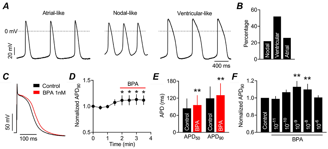
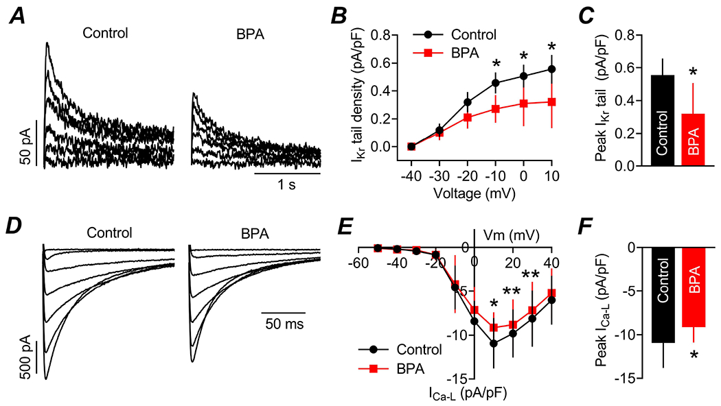
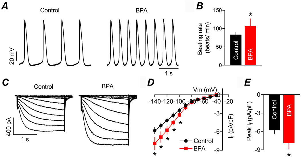
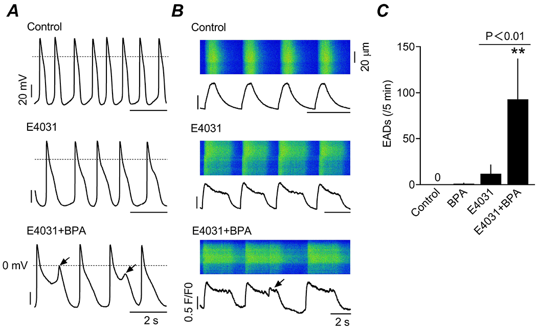
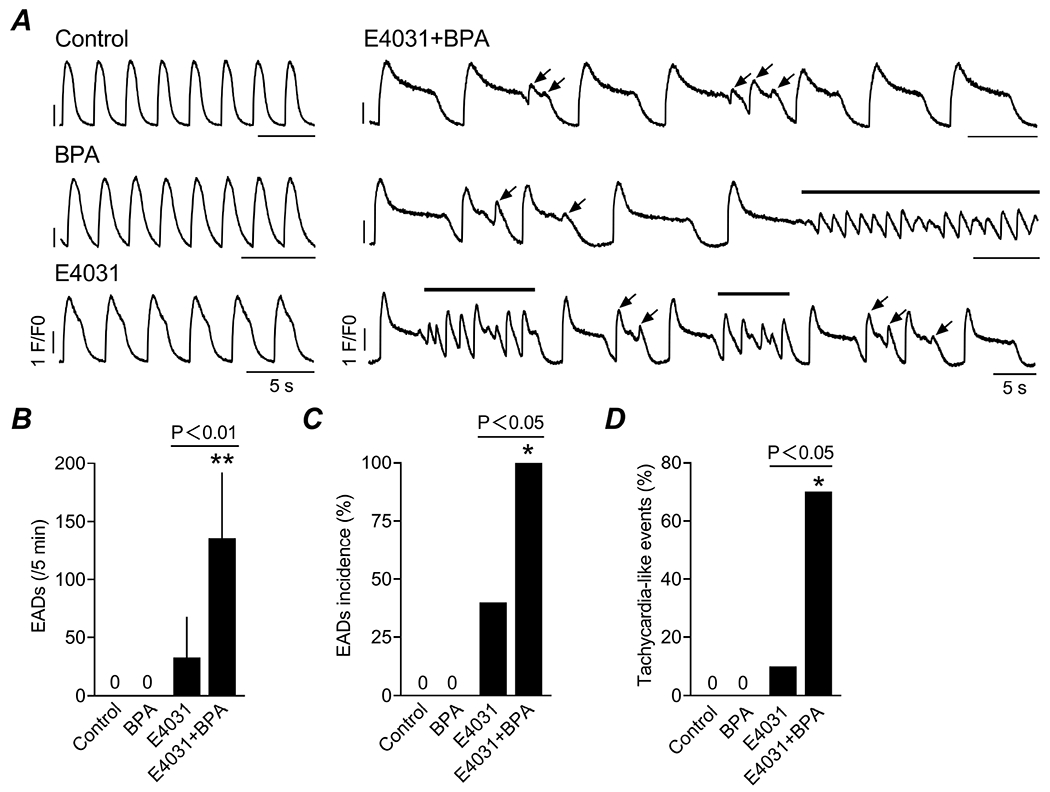
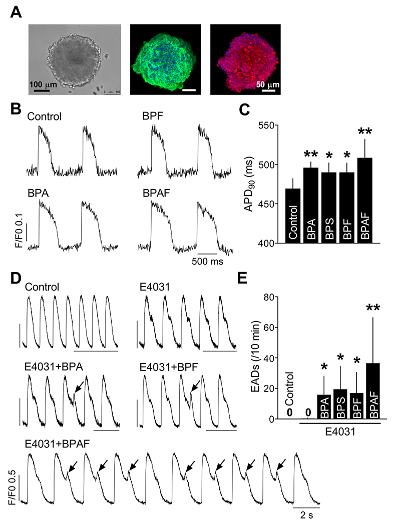
Bisphenol A (BPA) and its analogs are common environmental chemicals with many potential adverse health effects. The impact of environmentally relevant low dose BPA on human heart, including cardiac electrical properties, is not understood. Perturbation of cardiac electrical properties is a key arrhythmogenic mechanism. In particular, delay of cardiac repolarization can cause ectopic excitation of cardiomyocytes and malignant arrhythmia. This can occur as a result of genetic mutations (i.e., long QT (LQT) syndrome), or cardiotoxicity of drugs and environmental chemicals. To define the impact of low dose BPA on electrical properties of cardiomyocytes in a human-relevant model system, we examined the rapid effects of 1 nM BPA in human induced pluripotent stem cell-derived cardiomyocytes (hiPSC-CMs) using patch-clamp and confocal fluorescence imaging. Acute exposure to BPA delayed repolarization and prolonged action potential duration (APD) in hiPSC-CMs through inhibition of the hERG K+ channel. In nodal-like hiPSC-CMs, BPA acutely increased pacing rate through stimulation of the If pacemaker channel. Existing arrhythmia susceptibility determines the response of hiPSC-CMs to BPA. BPA resulted in modest APD prolongation but no ectopic excitation in baseline condition, while rapidly promoted aberrant excitations and tachycardia-like events in myocytes that had drug-simulated LQT phenotype. In hiPSC-CM-based human cardiac organoids, the effects of BPA on APD and aberrant excitation were shared by its analog chemicals, which are often used in "BPA-free" products, with bisphenol AF having the largest effects. Our results reveal that BPA and its analogs have repolarization delay-associated pro-arrhythmic toxicity in human cardiomyocytes, particularly in myocytes that are prone to arrhythmias. The toxicity of these chemicals depends on existing pathophysiological conditions of the heart, and may be particularly pronounced in susceptible individuals. An individualized approach is needed in risk assessment and protection.

Keywords: Arrhythmia; Bisphenol A; Human cardiac organoid; Human iPSC-CMs; Long QT syndrome.


Conflict of interest statement

Declaration of competing interest The authors declare that they have no known competing financial interests or personal relationships that could have appeared to influence the work reported in this paper.

Figures

Figure 1.
Figure 1.. Acute exposure to low dose BPA prolongs APD in hiPSC-CMs.
(A) Representative AP traces recorded from single hiPSC-CMs using perforated patch-clamp method; three typical types of AP morphology, atrial-, nodal- and ventricular-like, were observed. (B) Percentage of nodal-, ventricular-, and atrial-like cells in the examined hiPSC-CMs. (C) Representative AP traces recorded from ventricular-like hiPSC-CMs before (control) and after 1 nM BPA exposure (1-4 min). (D) Time course of AP duration change measured as 90% repolarization (APD90) following BPA exposure (n = 7). APD90 was normalized to control. (E) Average AP duration at 50% repolarization (APD50) and APD90 in hiPSC-CMs before and after BPA exposure (n = 7). (F) Dose-dependent effect of BPA on APD in hiPSC-CMs. APD90 in the presence of indicated concentrations of BPA were normalized to their respective controls. N = 5 myocytes for 10−11, 10−10 and 10−6 M, 6 for 10−8 M, and 7 for 10−9 M, respectively. *: P< 0.05; **: P< 0.01 vs control in paired t-test.
Figure 2.
Figure 2.. Low dose BPA rapidly inhibits Ikr and ICa-L in hiPSC-CMs.
(A) Representative current traces of IKr tail recorded from single ventricular-like hiPSC-CMs before (control) and after BPA exposure at 1 nM. IKr was elicited by 2-s depolarization steps ranging from −30 mV to 10 mV in 10 mV increments, from a holding voltage of −50 mV, and recorded at −40 mV. (B) Current-voltage (I-V) relationship of average IKr tail in hiPSC-CMs under control and upon exposure to BPA (n = 6). IKr tail current densities were plotted against the voltages of the depolarization steps. (C) Average peak Ikr tail density in hiPSC-CMs before and after BPA exposure (n = 6). (F) Representative traces of the L-type Ca2+ current (ICa-L) recorded from ventricular-like hiPSC-CMs before and after acute exposure to 1 nM BPA. ICa-L was elicited by 300 ms depolarizing voltage steps ranging from −50 mV to 40 mV in 10 mV increments, from a holding potential of −70 mV. (G) I-V relationship of ICa-L in hiPSC-CMs under control and upon exposure to BPA (n = 7). (H) Average peak ICa-L density in hiPSC-CMs before and after BPA exposure (n = 7). *: P< 0.05; **: P< 0.01 vs control in paired t-test.
Figure 3.
Figure 3.. Rapid exposure to BPA increases automaticity and If current in hiPSC-CMs.
(A) Representative AP traces recorded from spontaneously beating nodal-like single hiPSC-CMs before (control) and after BPA exposure at 1 nM. (B) Average beating rate (beats/min) in hiPSC-CMs under control and upon exposure to BPA (n = 4). (C) Representative traces of If current recorded from single hiPSC-CMs before and after 1 nM BPA exposure. If was elicited by 2-s hyperpolarizing pulses ranging from −40 mV to −140 mV in 10 mV increments, from a holding potential of −30 mV. (D) I-V relationship of If in hiPSC-CMs under control and upon exposure to BPA (n = 4). (E) Average peak If density in hiPSC-CMs before and after BPA exposure (n = 4). *: P< 0.05 vs control in paired t-test.
Figure 4.
Figure 4.. Effect of BPA on aberrant ectopic excitation in hiPSC-CMs with simulated LQT syndrome phenotype.
(A) Representative recordings of AP from spontaneously beating single hiPSC-CMs under control, 1 nM BPA or 10 nM E4031 alone, and BPA in the presence of E4031. 10 nM E4031 partially blocks IKr and simulates LQT syndrome type 2 phenotype (LQT2). Arrows indicate EADs. (B) Representative confocal line-scan images of Ca2+ transients in beating hiPSC-CMs and corresponding line plots of Ca2+ fluorescence intensity under control, BPA or E4031 alone, and BPA in the presence of E4031. Arrows: spontaneous ectopic excitations indictive of EADs. (C) Average frequency of EADs in hiPSC-CMs under various treatments. N = 10 myocytes for Control, 8 for both BPA and E4031+BPA, and 6 for E4031, respectively. F/F0, fluorescence intensity ratio. **: P< 0.01 vs control in one-way ANOVA.
Figure 5.
Figure 5.. Effect of BPA on arrhythmic events in hiPSC-CM monolayers with simulated LQT syndrome phenotype.
(A) Representative recordings of Ca2+ transients from beating hiPSC-CM monolayers under control, 1 nM BPA or 10 nM E4031 alone, and BPA in the presence of E4031. Arrows and thick lines indicate EADs and tachycardia-like arrhythmic events, respectively. (B) Average frequency of EADs in hiPSC-CM monolayers under various treatments (n= 10 monolayers/treatment). (C) Incidence of EADs in hiPSC-CM monolayers under various treatments (n= 10 monolayers/treatment). (D) Incidence of tachycardia-like arrhythmic events in hiPSC-CM monolayers under various treatments (n= 10 monolayers/treatment). F/F0, fluorescence intensity ratio. *: P< 0.05; **: P< 0.01 vs control in one-way ANOVA or chi-square test.
Figure 6.
Figure 6.. Effects of BPA and related bisphenol analogs on APD and EAD arrhythmia events in human cardiac organoids.
(A) Representative brightfield and immunofluorescence images of human cardiac organoids. Left, brightfield images of organoids. Center and right, immunofluorescence images of organoids stained for cardiomyocyte-specific markers cardiac troponin T (green) and sarcomeric α-actinin (red). DAPI (blue): nuclear counterstain. (B) Representative AP traces from organoids under control and indicated acute bisphenol exposure. F/F0, fluorescence intensity ratio. (C) Average APD90 in organoids under various treatments (n = 10 organoids for control and 7 for all bisphenol treatments). (D) Representative recordings of Ca2+ transients from organoids under control, 10 nM E4031 alone, and indicated bisphenols in the presence of E4031. Arrows indicate EADs. (E) Average frequency of EADs in organoids under various treatments (n = 11 organoids for control and 8 for all treatment groups). The concentration used for all bisphenols was 1 nM. *: P< 0.05; **: P< 0.01 vs control in one-way ANOVA.

Similar articles

Cited by

References

    1. Antzelevitch C, Burashnikov A, 2011. Overview of Basic Mechanisms of Cardiac Arrhythmia. Card Electrophysiol Clin 3, 23–45. - PMC - PubMed
    1. Bezzina CR, Lahrouchi N, Priori SG, 2015. Genetics of sudden cardiac death. Circ Res 116, 1919–1936. - PubMed
    1. Brette F, Shiels HA, Galli GL, Cros C, Incardona JP, Scholz NL, Block BA, 2017. A Novel Cardiotoxic Mechanism for a Pervasive Global Pollutant. Sci Rep 7, 41476. - PMC - PubMed
    1. Cai S, Rao X, Ye J, Ling Y, Mi S, Chen H, Fan C, Li Y, 2020. Relationship between urinary bisphenol a levels and cardiovascular diseases in the U.S. adult population, 2003-2014. Ecotoxicol Environ Saf 192, 110300. - PubMed
    1. Calafat AM, Kuklenyik Z, Reidy JA, Caudill SP, Ekong J, Needham LL, 2005. Urinary concentrations of bisphenol A and 4-nonylphenol in a human reference population. Environ Health Perspect 113, 391–395. - PMC - PubMed

MeSH terms

LinkOut - more resources