. 2023 May 15;63(5):179-190.
doi: 10.2176/jns-nmc.2022-0271. Epub 2023 Mar 31.

Clinical Impacts of Stereotactic Electroencephalography on Epilepsy Surgery and Associated Issues in the Current Situation in Japan

Affiliations

Clinical Impacts of Stereotactic Electroencephalography on Epilepsy Surgery and Associated Issues in the Current Situation in Japan

Satoshi Maesawa et al. Neurol Med Chir (Tokyo). .

Abstract

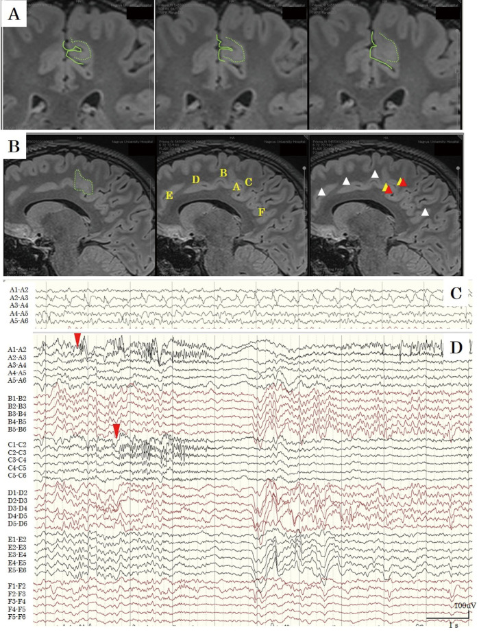
Stereotactic electroencephalography (SEEG) is receiving increasing attention as a safe and effective technique in the invasive evaluation for epileptogenic zone (EZ) detection. The main clinical question is whether the use of SEEG truly improves outcomes. Herein, we compared outcomes in our patients after three types of intracranial EEG (iEEG): SEEG, the subdural electrode (SDE), and a combined method using depth and strip electrodes. We present here our preliminary results from two demonstrative cases. Several international reports from large epilepsy centers found the following clinical advantages of SEEG: 1) three-dimensional analysis of structures, including bilateral and multilobar structures; 2) low rate of complications; 3) less pneumoencephalopathy and less patient burden during postoperative course, which allows the initiation of video-EEG monitoring immediately after implantation and does not require resection to be performed in the same hospitalization; and 4) a higher rate of good seizure control after resection. In other words, SEEG more accurately identified the EZ than the SDE method. We obtained similar results in our preliminary experiences under limited conditions. In Japan, as of August 2022, dedicated electrodes and SEEG accessories have not been approved and the use of the robot arm is not widespread. The Japanese medical community is hopeful that these issues will soon be resolved and that the experience with SEEG in Japan will align with that of large epilepsy centers internationally.

Keywords: complication; epilepsy surgery; focal epilepsy; outcomes; stereotactic electroencephalography (SEEG).


Conflict of interest statement

All authors declare that there are no conflicts of interest (COIs) regarding this article according to the criteria of The Japan Neurosurgical Society. They have completed the self-reported registration of their COI status to the society.

Figures

Fig. 1
Fig. 1
Preoperative images and anatomo-electro-clinical correlation (AEC) hypothesis for Case 1. A: A fluid attenuated inversion recovery image shows a partial defect in the opercular part of the right inferior frontal gyrus. B: 18F-fluorodeoxyglucose positron emission tomography shows low uptake of glucose around this area. C: Schematic images for AEC hypothesis. In the first hypothesis (red line), the seizure could start from the lesion in the opercular part. Impaired awareness could be associated with the anterior cingulate cortex or the orbitofrontal cortex. In the second hypothesis, seizure onset is the insula because the patient reported occasionally experiencing vague dysesthesia. Propagation to the supplementary motor area may cause fencing posture.
Fig. 2
Fig. 2
A: Electrode location and ictal recording of stereotactic electroencephalography (SEEG). Tract A: around the lesion in the opercular part in the inferior frontal gyrus, Tract B: the orbitofrontal cortex to the rectal gyrus, Tract C: frontopolar part to the rostral anterior cingulate cortex (ACC), Tract D: dorsolateral prefrontal cortex to the rostral ACC, Tract E: premotor cortex to the central cingulate cortex, Tract F: the supplementary motor area, Tract G: triangular part, Tract H: primary motor cortex, and Tract I: anterior insula. Tracts B, F, and I were inserted in the oblique fashion, and the other tracts were made in the orthogonal fashion. We confirmed targets and tracts (yellow) using superimposed images of postoperative computed tomography (CT) and preoperative magnetic resonance imaging. White dots are the contacts of the electrodes, observed on postoperative CT. All electrodes were accurately inserted according to the preoperative plans. B: In an ictal recording of SEEG, after a high-amplitude spike appeared in the wide area, a low-amplitude fast wave in the triangular part (G1, G2) began. This activity propagated to the premotor cortex (E5, E6).
Fig. 3
Fig. 3
The appearance of stereotactic electroencephalography (SEEG) in Case 2. A: Fluid attenuated inversion recovery images show blurring in the border between the gray and white matter and a high-intensity area in the thickened cortex in the paracentral lobule, extending to the premotor area in the left medial frontal lobe (contoured by a green dot line). B: The electrode position of SEEG: (A) High-intensity lesion in the paracentral lobule; (B) middle cingulate cortex; (C) superior side of the lesion; (D) anterior cingulate cortex; (E) rostral side of the frontal lobe; and (F) posterior cingulate gyrus. The yellow triangle represents the seizure onset zone. The red triangle represents the electrodes that reproduced her habitual seizure by stimulation. The white triangle represents no response of the electrodes to stimulation. C: Frequent interictal spikes in A2 and A3, consistent with the high-intensity lesion. D: Interictal recording demonstrating that the seizure onset zone was in the lesion (A2 and A3), followed by the dorsal side of the paracentral gyrus (C5 and C6).

Similar articles

Cited by

References

    1. Bancaud J, Angelergues R, Bernouilli C, et al. : Functional stereotaxic exploration (SEEG) of epilepsy. Electroencephalogr Clin Neurophysiol 28: 85-86, 1970 - PubMed
    1. Cardinale F, Cossu M, Castana L, et al. : Stereoelectroencephalography: Surgical methodology, safety, and stereotactic application accuracy in 500 procedures. Neurosurgery 72: 353-366, 2013 - PubMed
    1. González-Martínez J, Bulacio J, Thompson S, et al. : Technique, results, and complications related to robot-assisted stereoelectroencephalography. Neurosurgery 78: 169-180, 2016 - PubMed
    1. Talairach J, Bancaud J: Stereotaxic approach to epilepsy methodology of anatomo-functional stereotaxic investigations. Prog Neurol Surg Basel 297-354, 1973
    1. Bancaud J: Surgery of epilepsy based on stereotactic investigations–the plan of the SEEG investigation. Acta Neurochir Suppl 30: 25-34, 1980 - PubMed