

A rare case report of tenosynovial chondromatosis of the semimembranosus-medial collateral ligament bursa
- PMID: 37005684
- PMCID: PMC10067226
- DOI: 10.1186/s12891-023-06337-6
A rare case report of tenosynovial chondromatosis of the semimembranosus-medial collateral ligament bursa
Abstract
Background: Synovial chondromatosis is an uncommon metaplastic process of the synovial lining that results in the formation of cartilaginous nodules within joints or their associated bursae or tendon sheaths. Radiologic evidence of mineralized bodies within these structures is typically pathognomonic for this condition. Extraarticular chondromatosis is rarer than intraarticular chondromatosis, and the knee is affected less frequently than the smaller joints of the hands and feet. To our knowledge, no reports describing this condition in the semimembranosus-medial collateral ligament (SM-MCL) bursa have been published.
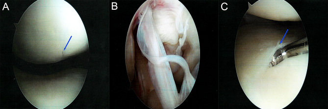
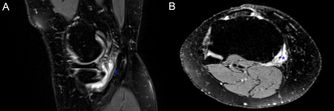
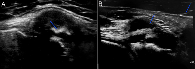
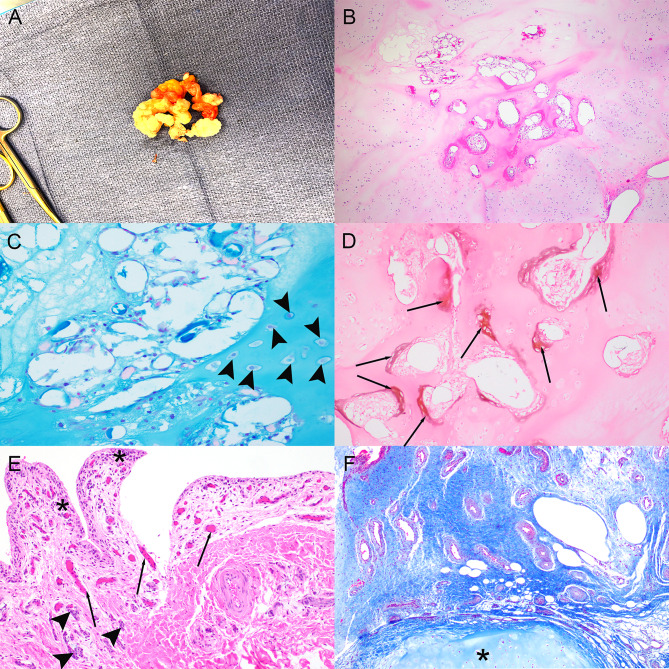
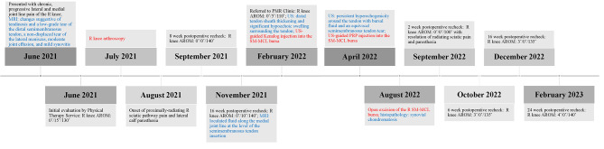
Case presentation: We describe a case of tenosynovial chondromatosis in a 37-year-old woman. The case was atypical for both the location within the SM-MCL bursa and the paucity of radiodense or hypointense changes to support a clinical suspicion of chondroid metaplasia on radiographs and T2-weighted MRI, respectively. Recreational weightlifting and swimming by the patient were impaired by chronic pain, and restricted range of motion of the ipsilateral knee persisted despite extensive skilled physical therapy and injections of both corticosteroids and platelet-rich plasma. Thirteen months after a diagnostic and therapeutic knee arthroscopy, open surgical excision of the SM-MCL bursal body was performed, and knee pain and range of motion improved by the 6-week postoperative reevaluation. Pathologic evaluation of the excised tissue was consistent with tenosynovial chondromatosis.
Conclusions: Synovial chondromatosis should be considered in the differential diagnosis for recalcitrant bursitis, even in the absence of classic imaging findings.
Keywords: Knee; Semimembranosus-medial collateral ligament bursa; Tenosynovial chondromatosis.
© 2023. The Author(s).
Conflict of interest statement
The authors declare that they have no competing interests.
Figures
References
-
- De Maeseneer M, Marcelis S, Boulet C, Kichouh M, Shahabpour M, de Mey J, et al. Ultrasound of the knee with emphasis on the detailed anatomy of anterior, medial, and lateral structures. Skeletal Radiol. 2014 Aug;43(8):1025–39. - PubMed
-
- Hennigan SP, Schneck CD, Mesgarzadeh M, Clancy M. The semimembranosus-tibial collateral ligament bursa. Anatomical study and magnetic resonance imaging. J Bone Joint Surg Am. 1994 Sep;76(9):1322–7. - PubMed
-
- Rothstein CP, Laorr A, Helms CA, Tirman PF. Semimembranosus-tibial collateral ligament bursitis: MR imaging findings. AJR Am J Roentgenol. 1996 Apr;166(4):875–7. - PubMed
-
- Saavedra M, Navarro-Zarza JE, Villaseñor-Ovies P, Canoso JJ, Vargas A, Chiapas-Gasca K, et al. Clinical anatomy of the knee. Reumatol Clin. 2012 Jan;8(Suppl 2):39–45. - PubMed
-
- Vicentini JRT, Chang CY. MR imaging of the knee bursae and bursal pathology. Magn Reson Imaging Clin N Am. 2022 May;30(2):241–60. - PubMed
Publication types
MeSH terms
LinkOut - more resources
Full Text Sources
