. 2023 Apr 1;14(1):56.
doi: 10.1186/s13244-023-01410-z.

CT appearances and classification of hepatic epithelioid hemangioendothelioma

Affiliations

CT appearances and classification of hepatic epithelioid hemangioendothelioma

Haidong Tan et al. Insights Imaging. .

Abstract

Background: Hepatic epithelioid hemangioendothelioma (HEH) is extremely rare, and CT features have never been analyzed in a large group of patients.

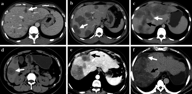
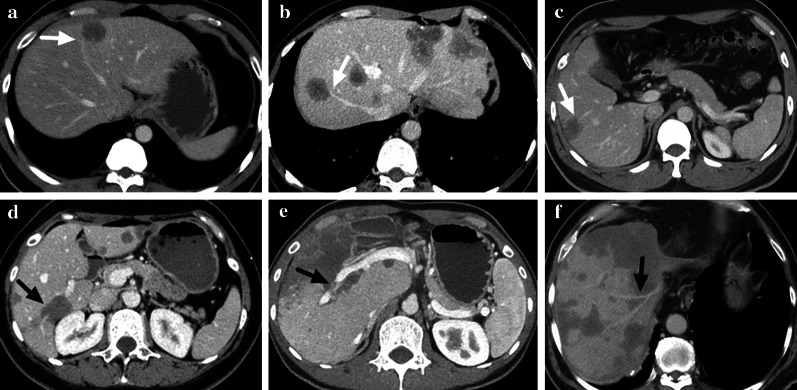
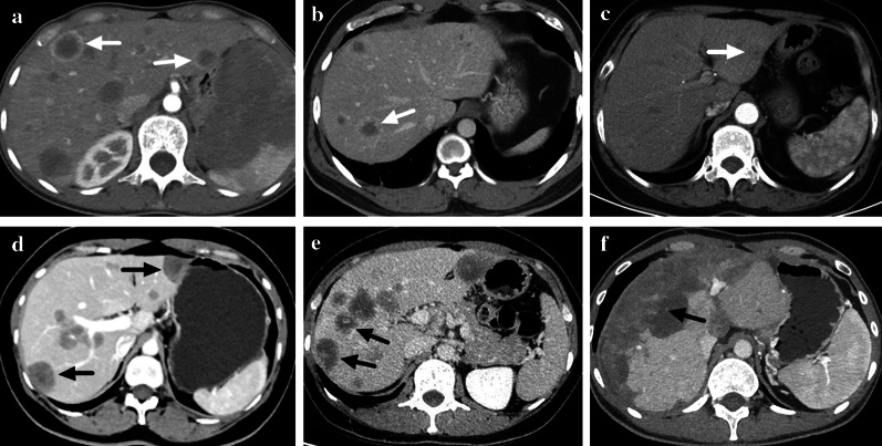
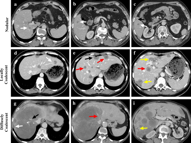
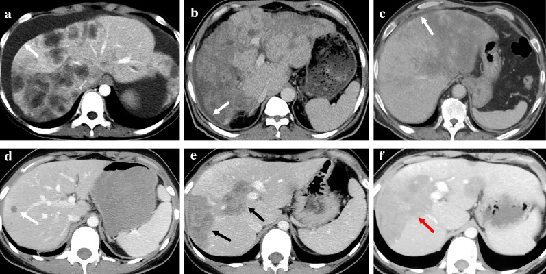
Methods: A retrospective study was designed to review the contrast-enhanced CT images of HEH patients. Intrahepatic lesions were categorized into three types: nodular, locally coalescent (coalescent lesion contained in one segment) or diffusely coalescent (coalescent lesion occupied more than one segment). CT features were compared among lesions of different sizes and patients with different lesion types.

Results: A total of 93 HEH patients were included in this study, and 740 lesions were analyzed. The results of per-lesion analysis showed that medium lesions (2-5 cm) had the highest rate of lollipop sign (16.8%) and target-like enhancement (43.1%), while lesions in large group (> 5 cm) had the highest rate of capsular retraction (38.8%) and vascular invasion (38.8%). The differences on enhancement pattern and the rates of lollipop sign and capsular retraction were significant among lesions of different sizes (p < 0.001, respectively). The results of per-patient analysis showed that patients in locally coalescent group had the highest rates of lollipop sign (74.3%) and target sign (94.3%). All patients in diffusely coalescent group had capsular retraction and vascular invasion. CT appearances of capsular retraction, lollipop sign, target sign and vascular invasion differed significantly among patients with different lesion types (p < 0.001, p = 0.005, p = 0.006 and p < 0.001, respectively).

Conclusion: CT features variated among HEH patients with different lesion types, and radiological appearances of HEH should be classified into nodular type, locally coalescent type and diffusely coalescent type.

Keywords: CT; Hepatic epithelioid hemangioendothelioma; Hepatic tumors; Rare liver tumors.


Conflict of interest statement

The authors declare that they have no conflict of interest.

Figures

Fig. 1
Fig. 1
Patient flow chart for inclusion. A total of 135 pathologically confirmed hepatic epithelioid hemangioendothelioma (HEH) patients were regularly followed up in our center. Excluded were 25 patients with no CT scan within 30 days prior to surgery or biopsy, 10 patients with incomplete CT scan, 5 patients with CT images of insufficient quality and 2 patients with severe hepatic steatosis
Fig. 2
Fig. 2
a A nodular lesion with target sign was marked with a white arrow. b A locally coalescent lesion with target sign was marked with a white arrow. c A diffusely coalescent lesion occupied multiple liver segments (marked with a white arrow). Target sign could also be found in the lesion (marked with a black arrow). d Target sign could be found on unenhanced CT scan (marked with a white arrow). f Capsular retraction caused by a subcapsular lesion could be detected (marked with a black arrow). e A diffusely coalescent lesion (marked with a black arrow) was accompanied with capsular retraction and perihepatic effusion (marked with a white arrow)
Fig. 3
Fig. 3
a–c Lollipop sign on portal phase of three HEH patients, which showed vessels terminated at the edge of a well-defined lesion (marked with white arrows). d–f Vascular invasion on portal phase of three HEH patients, which showed vessels penetrated lesions or was surrounded by lesions with or without intact vessel structure (marked with black arrows)
Fig. 4
Fig. 4
Enhancement patterns of HEH lesions on contrast-enhanced CT scan. a Ring-like enhancement on arterial phase (marked with white arrows). b Ring-like enhancement on portal phase (marked with a white arrow). c Target-like enhancement on arterial phase (marked with a white arrow). d Target-like enhancement on portal phase (marked with black arrows). e Core enhancement on portal phase (marked with black arrows). f Heterogenous enhancement on portal phase (marked with a black arrow)
Fig. 5
Fig. 5
a–c Contrast-enhanced CT scan of a 44-year-old male HEH patient in nodular group. Multiple intrahepatic nodular lesions could be found (marked with white arrows) with one subcapsular lesion (marked with a black arrow). No target sign or lollipop sign was detected. d–f Contrast-enhanced CT scan of a 32-year-old female HEH patient in locally coalescent group. Locally coalescent lesions could be found (marked with yellow arrows). A subcapsular lesion caused capsular retraction (marked with a black arrow). Both target sign (marked with red arrows) and lollipop sign (marked with a white arrow) could be detected. g–i Contrast-enhanced CT scan of a 68-year-old female HEH patient in diffusely coalescent group. Right hepatic vein disappeared, and middle hepatic vein was involved by the diffusely coalescent lesion (marked with a red arrow). Both capsular retraction (marked with a white arrow) and calcification (marked with a black arrow) could be detected. One subcapsular lesion with calcification and capsular retraction was marked with a yellow arrow
Fig. 6
Fig. 6
a–c Perihepatic effusion of three HEH patients on CT scan (marked with white arrows). d–f The changes of CT appearances of a 28-year-old male HEH patient. During the period of 6 years follow-up, the patient received no treatment. d At the time of diagnosis, nodular lesion could be found (marked with a white arrow). e The CT scan at 4 years after the diagnosis showed locally coalescent lesions (marked with black arrows). f The CT scan at 6 years after the diagnosis showed diffusely coalescent lesion (marked with a red arrow)

References

    1. Sardaro A, Bardoscia L, Petruzzelli MF, Portaluri M. Epithelioid hemangioendothelioma: an overview and update on a rare vascular tumor. Oncol Rev. 2014;8(2):259. - PMC - PubMed
    1. Chahrour MA, Khachfe HH, Habib JR, El-Asmar R, Saifi O, Jamali FR. Treatment and prognosis of hepatic epithelioid hemangioendothelioma: a SEER database analysis. World J Surg. 2021;45(9):2886–2894. doi: 10.1007/s00268-021-06165-6. - DOI - PubMed
    1. Onishi Y, Kusumoto M, Motoi N, et al. Natural history of epithelioid hemangioendothelioma of the liver: CT findings of 15 cases. Acad Radiol. 2021;28(6):778–782. doi: 10.1016/j.acra.2020.04.015. - DOI - PubMed
    1. Liu XL, Yang ZY. Outcomes of hepatic epithelioid hemangioendothelioma with different managements: a retrospective investigation. Eur Rev Med Pharmacol Sci. 2021;25(12):4274–4282. - PubMed
    1. Liu X, Zhang Z, Huang J, Tan H, Yang Z. Efficacy and safety of interferon-alpha 2b for patients with hepatic epithelioid hemangioendothelioma: outcomes of a case-series analysis. Cancer Manag Res. 2021;13:8273–8279. doi: 10.2147/CMAR.S334171. - DOI - PMC - PubMed

LinkOut - more resources