. 2023 Mar 15:14:1139123.
doi: 10.3389/fimmu.2023.1139123. eCollection 2023.

Mapping the SLP76 interactome in T cells lacking each of the GRB2-family adaptors reveals molecular plasticity of the TCR signaling pathway

Affiliations

Mapping the SLP76 interactome in T cells lacking each of the GRB2-family adaptors reveals molecular plasticity of the TCR signaling pathway

Kilian Ruminski et al. Front Immunol. .

Abstract

The propagation and diversification of signals downstream of the T cell receptor (TCR) involve several adaptor proteins that control the assembly of multimolecular signaling complexes (signalosomes). The global characterization of changes in protein-protein interactions (PPI) following genetic perturbations is critical to understand the resulting phenotypes. Here, by combining genome editing techniques in T cells and interactomics studies based on affinity purification coupled to mass spectrometry (AP-MS) analysis, we determined and quantified the molecular reorganization of the SLP76 interactome resulting from the ablation of each of the three GRB2-family adaptors. Our data showed that the absence of GADS or GRB2 induces a major remodeling of the PPI network associated with SLP76 following TCR engagement. Unexpectedly, this PPI network rewiring minimally affects proximal molecular events of the TCR signaling pathway. Nevertheless, during prolonged TCR stimulation, GRB2- and GADS-deficient cells displayed a reduced level of activation and cytokine secretion capacity. Using the canonical SLP76 signalosome, this analysis highlights the plasticity of PPI networks and their reorganization following specific genetic perturbations.

Keywords: GRB2-family adaptors; SLP76; T cell signaling; TCR; mass spectrometry.


Conflict of interest statement

The authors declare that the research was conducted in the absence of any commercial or financial relationships that could be construed as a potential conflict of interest.

Figures

Figure 1
Figure 1
Generation and validation SLP76OST Jurkat cells deficient in GRB2-family adaptors. (A) Immunoblot analysis of equal amounts of lysates from parental Jurkat, SLP76OST (WT), SLP76OSTGADS-/- (GADS-/-), SLP76OSTGRB2-/- (GRB2-/-) and SLP76OSTGRAP-/- (GRAP-/-) cells probed with the indicated antibodies. (B) Expression of CD6, CD3 and CD28 at the surface of parental and Jurkat variants indicated in (A), analyzed using flow cytometry. Data are representative of at least three independent experiments.
Figure 2
Figure 2
GRB2-family adaptors dependent and independent SLP76 protein interactions. (A) Volcano plot showing protein enrichment of affinity purified SLP76OST from WT Jurkat cells compared to parental Jurkat cells expressing similar levels of untagged SLP76 proteins at 60 s after CD3 stimulation. Enriched preys are identified by a fold change > 3 and pvalue <0.01. Canonical SLP76 interactors are highlighted in red. (B) Dot plot showing the interaction stoichiometry of SLP76 with the 23 high-confidence preys for which the interaction stoichiometry is regulated by the inactivation of one the GRB2-family adaptors (fold change > 2.5 and pvalue <0.05). The interaction stoichiometry has been row-normalized to its maximum value observed over stimulation conditions (see key for Normalized Stoichiometry). The pvalue key is related to protein enrichment of the analysis in (A). GRB2-family adaptors are framed in black. The two distinct SLP76 modules are framed in blue and green.
Figure 3
Figure 3
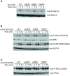
Analysis of TCR-induced molecular events in cells deficient in GRB2-family adaptors. (A) WT, GADS-/-, GRB2-/- and GRAP-/- Jurkat cells were left unstimulated (-) or stimulated for 1 or 5 min with anti-CD3 antibody. Equal amounts of protein lysates were analyzed by immunoblot with phospho-specific antibodies for tyrosine motifs (P-Tyr), ERK1/2 (T202/Y204), SLP76 (Y128), PLCγ1 (Y783) and LAT (Y191). VAV1 and total proteins serve as a loading control (left and right panel respectively). The CD6 expression was also assessed for each Jurkat variants. (B–E) The same cells as in (A) were left unstimulated (-) or stimulated (+) with anti-CD3 antibody and subsequently lysed. Equal amounts of cell lysates were incubated with the specified antibodies (IP) and the resulting immunoprecipitates analyzed by immunoblot with the antibodies specified in the left margin (IB). Data are representative of at least two independent experiments.
Figure 4
Figure 4
Impaired T cell functions in GADS and GRB2 deficient cells. (A) WT, GADS-/-, GRB2-/- and GRAP-/- Jurkat cells were loaded with Indo-1AM and stimulated with 2 or 0.5 μg of anti-CD3 antibody (final concentrations 5 μg/ml and 1.25 μg/ml respectively) and concentration of intracellular calcium was monitored for 5 min at 37°C by flow cytometry. The TCR-independent response was assessed by addition of Ionomycin (Iono) after 4 min. (B) WT and Jurkat variants were stimulated with Raji cells in presence of increasing amount of SEE. The expression of CD69 was assessed 24 h after stimulation by flow cytometry. A graph of percentage of CD69 positive cells was plotted as a function of SEE concentrations. (C) ELISA analysis of IL-2 production by the specified cells 24 h after stimulation with Raji cells and SEE. (mean +/- sd of technical replicates). Data are representative of at least three independent experiments.
Figure 5
Figure 5
Normal co-stimulatory signaling in Jurkat cells deficient in GRB2-family adaptors. (A) WT, GADS-/-, GRB2-/- and GRAP-/- Jurkat cells were left unstimulated (-) or stimulated for 2 or 5 min with anti-CD6 antibody. Equal amounts of protein lysates were analyzed by immunoblot with phospho-specific ERK1/2 antibody and with anti-ERK1/2 antibody. (B) Same cells as in A were left unstimulated (-) or stimulated for 2, 5 or 10 min with anti-CD3 and anti-CD28 antibodies. Equal amounts of protein lysates were analyzed by immunoblot with indicated phospho-specific antibodies and loading control was assessed with anti-SLP76 antibody. (C) Analysis of the PKCθ phosphorylation in cells and experimental conditions described in (B). Data are representative of at least two independent experiments.
Figure 6
Figure 6
Preserved ITK-SLP76 interaction in the absence of GRB2-family adaptors. (A) The abundances of ITK was estimated in SLP76OST AP-MS from cells that were left unstimulated (0) or stimulated for 1 or 5 min with anti-CD3 antibody in three independent biological replicates (R1, R2 and R3). (B) Comparison of ITK-SLP76 interaction stoichiometry in cells stimulated with anti-CD3 for 1 min. Imputed missing values are represented with lighter shaded dots. (C) MS intensities of specified phosphosites detected on the SLP76OST molecules in experimental conditions described in (A) WT refers to the SLP76OST variant while GADS-/-, GRB2-/- and GRAP-/- refer to the SLP76OST variants deficient for GADS (GRAP2), GRB2 and GRAP respectively. Normalized intensities (see Materials and Methods) from parental Jurkat (SLP76) and SLP76OST cells were compared using a two-sided Welch t-test (symbols used according to the t-test P-value: N.S., P > 0.05; *P ≤ 0.05; **P ≤ 0.01.

Similar articles

Cited by

References

    1. Gaud G, Lesourne R, Love PE. Regulatory mechanisms in T cell receptor signalling. Nat Rev Immunol (2018) 18(8):485–97. doi: 10.1038/s41577-018-0020-8 - DOI - PubMed
    1. Jordan MS, Koretzky GA. Coordination of receptor signaling in multiple hematopoietic cell lineages by the adaptor protein SLP-76. Cold Spring Harb Perspect Biol (2010) 2(4):a002501. doi: 10.1101/cshperspect.a002501 - DOI - PMC - PubMed
    1. Yablonski D. Bridging the gap: Modulatory roles of the Grb2-family adaptor, gads, in cellular and allergic immune responses. Front Immunol (2019) 10:1704. doi: 10.3389/fimmu.2019.01704 - DOI - PMC - PubMed
    1. Buday L, Egan SE, Rodriguez Viciana P, Cantrell DA, Downward J. A complex of Grb2 adaptor protein, sos exchange factor, and a 36-kDa membrane-bound tyrosine phosphoprotein is implicated in ras activation in T cells. J Biol Chem (1994) 269(12):9019–23. doi: 10.1016/S0021-9258(17)37070-9 - DOI - PubMed
    1. Balagopalan L, Coussens NP, Sherman E, Samelson LE, Sommers CL. The LAT story: a tale of cooperativity, coordination, and choreography. Cold Spring Harb Perspect Biol (2010) 2(8):a005512. doi: 10.1101/cshperspect.a005512 - DOI - PMC - PubMed

Publication types

MeSH terms

Substances