Review
. 2023 Mar 2;3(3):628-656.
doi: 10.1021/jacsau.2c00569. eCollection 2023 Mar 27.

Glycosaminoglycans: What Remains To Be Deciphered?

Affiliations
Review

Glycosaminoglycans: What Remains To Be Deciphered?

Serge Perez et al. JACS Au. .

Abstract

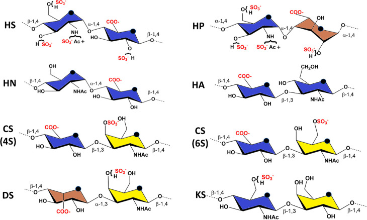
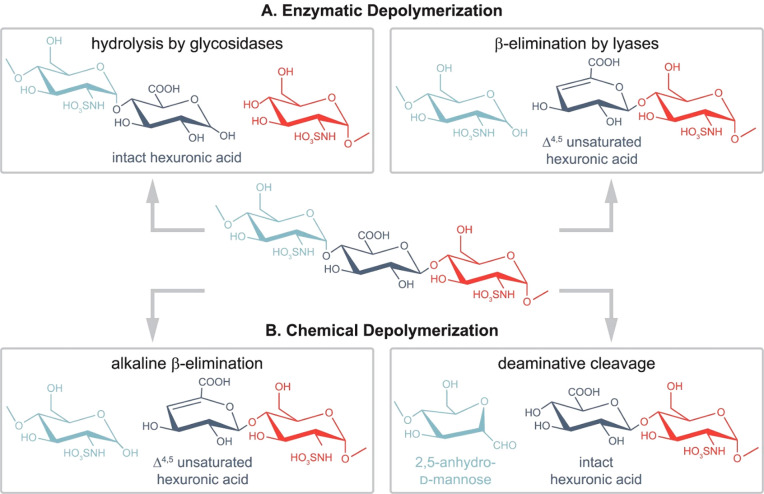
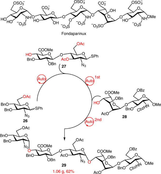
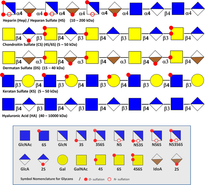
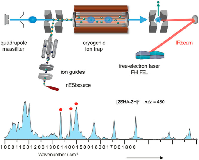
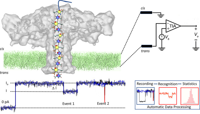
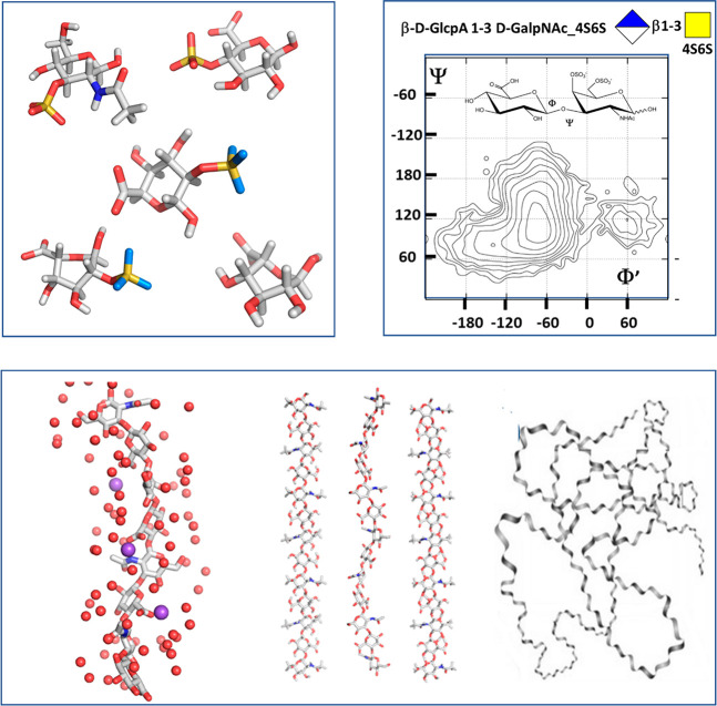
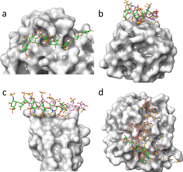
Glycosaminoglycans (GAGs) are complex polysaccharides exhibiting a vast structural diversity and fulfilling various functions mediated by thousands of interactions in the extracellular matrix, at the cell surface, and within the cells where they have been detected in the nucleus. It is known that the chemical groups attached to GAGs and GAG conformations comprise "glycocodes" that are not yet fully deciphered. The molecular context also matters for GAG structures and functions, and the influence of the structure and functions of the proteoglycan core proteins on sulfated GAGs and vice versa warrants further investigation. The lack of dedicated bioinformatic tools for mining GAG data sets contributes to a partial characterization of the structural and functional landscape and interactions of GAGs. These pending issues will benefit from the development of new approaches reviewed here, namely (i) the synthesis of GAG oligosaccharides to build large and diverse GAG libraries, (ii) GAG analysis and sequencing by mass spectrometry (e.g., ion mobility-mass spectrometry), gas-phase infrared spectroscopy, recognition tunnelling nanopores, and molecular modeling to identify bioactive GAG sequences, biophysical methods to investigate binding interfaces, and to expand our knowledge and understanding of glycocodes governing GAG molecular recognition, and (iii) artificial intelligence for in-depth investigation of GAGomic data sets and their integration with proteomics.


Conflict of interest statement

The authors declare no competing financial interest.

Figures

Figure 1
Figure 1
Principle constituent disaccharide units: HS, heparan sulfate: -4-D-GlcNAc-α1,4-D-GlcA-β1-. HP, heparin: -4-D-GlcN, NS, 6S-α1,4-L-IdoA-2S- α1-. HN, heparosan: -4-D-GlcNAc-α1,4-D-GlcA-β1-. HA, hyaluronan: 4-D-GlcA-β1-3-D-GlcNAc-β1. CS, (4S)-chondroitin 4-sulfate: 4-D-GlcA-β1-3-D-GalNAc, 4S-β1. CS, (6S)-chondroitin 6-sulfate: 4-D-GlcA-β1-3-D-GalNAc, 6S-β1. DS, dermatan sulfate: -4-L-IdoA-α1-3-D-GalNAc,4S-β1-. KS, keratan sulfate: -4-D-GlcNAc, 6S-β1-3-D-Gal-β1-. Color displayed in the monosaccharide units follows the SNFG recommendations.
Figure 2
Figure 2
Common glycosaminoglycan depolymerization strategies are illustrated through the example of heparan sulfate/heparin. (A, left) When glycosidases are used to depolymerize GAG chains, the resulting cleavage preserves the hexuronic acid stereochemistry. Enzymes with endolytic activity are required to obtain oligosaccharides covering the full sequence. As for moderately sulfated HS/heparin chains, using heparanase as endo β-glucuronidases cleaves at the reducing end of GlcA residues (A, right). Prokaryotic lyases, such as heparinase I–III, follow a β-eliminative mechanism, resulting in Δ4,5-unsaturated uronic acid residues at the new nonreducing end. Consequently, stereochemical information is lost in the process (B, left). Benzyl esterification with alkaline β-elimination mimics lyase activity and creates Δ4,5-unsaturated uronic acid residues at the new nonreducing end (B, right). While preserving hexuronic acid stereochemical information at the cleavage site, deamination cleavage alters the structure of the glucosamine through the formation of 2,5-anhydromannose. N-Acetyl groups on glucosamines block the reaction, making prior deacetylation necessary. Reproduced from ref (3). Copyright 2022, American Chemical Society.
Figure 3
Figure 3
Gram-scale synthesis of a protected fondaparinux pentasaccharide using a dual-mode automated solution-phase glycan synthesizer. A library of oligosaccharides covering various glycoforms and glycosidic linkages assembles rapidly, either in a general promoter-activation mode or light-induced-activation mode. The synthesis used thioglycoside d-glucuronic acid containing disaccharide with 3,6-di-O-acetyl groups for the α-directing glycosylation and l-iduronic-acid-containing disaccharide as building blocks. All compounds are readily obtained from commercially available monosaccharide or disaccharide intermediates. Reproduced with permission form ref (31). Copyright 2022, Springer Nature.
Figure 4
Figure 4
General structures of GAGs with their monosaccharide components. Overview of the characteristic monosaccharide components, N- and O-sulfation motifs, and linkages displayed per the SNFG representation. Repetitive hyaluronan chains (HA) are not modified by sulfation and epimerization. Chondroitin sulfate (CS), dermatan (DS), and keratan sulfate (KS) sulfate display a variety of sulfation motifs. Heparin and heparan sulfate represent the most diverse family of GAGs.
Figure 5
Figure 5
Schematic diagram of an IRMS instrument. The fragment ion charge ratio is measured via time-of-flight analysis. Helium droplets pick up trapped ions, which are immediately cooled to 0.37 K. There is subsequent irradiation of the droplets with monochromatic, high-intensity IR radiation, for example, using a free-electron laser (FEL). Reproduced with permission from ref (71). Copyright 2021, John Wiley and Sons.
Figure 6
Figure 6
Nanopore sequencing. Illustration of the organization of the translocation device showing the insertion of the aerolysin nanopore within the membrane and the elusive depiction of a chondroitin sulfate glycosaminoglycan passing through the channel of aerolysin. Nanopore experiments use a horizontal lipid bilayer Teflon device. The setup comprises the cis and trans chambers connected by a sub-millimeter inner diameter capillary. The lipid bilayer is formed by depositing a film of 1,2-diphytanoyl-sn-glycero-3-phosphocholine over a conical aperture of 20–30 μmin diameter that separates the cis and trans chambers. Two Ag–AgCl electrodes are installed in the cis and trans chambers filled with 100 μL of a buffer allowing the application of a fixed voltage and measurement of the ionic current. The entire setup is placed within a grounded Faraday cage to shield it from electromagnetic interference electrically. Upon its translocation, the polysaccharide inside the channel blocks the current. For each translocation event, the block current, the open pore current and the duration of the event are recorded for further identification and statistical analysis. Adapted from ref (75) and is licensed under CC BY 4.0, https://figshare.com/articles/figure/Nanopores_GAGs_sequencing/19391822.
Figure 7
Figure 7
From local to global. From quantum mechanics to coarse-grained simulation, computational methods yield continuous descriptions of the structural features occurring over a wide range of dimensions. Local properties include (1) the description of the monosaccharide low energy conformation characterized by the ring puckers; (2) the potential energy surface computed as a function of the values of the glycosidic torsion angles displaying the occurrence of the low energy conformers and the conformational pathways between them; (3) the interactions of the GAGs chains with ions and water molecules, and the occurrence of the several low energy helical structures of the GAG chain; and (4) the monitoring of fluctuations in lengths and volumes in disordered states and characterization of the radius of gyration and persistence lengths.
Figure 8
Figure 8
GAG-binding to proteins: From “high selectivity” to “non-selectivity”, a continuous spectrum of interactions intermediate cases exists. (a) The panel displays a system in which unique interactions between sulfate groups of heparin and basic residues of antithrombin result from a highly selective recognition. (b) The GAG–protein mode of interactions exhibits a “moderate selectivity” where several binding modes occur throughout the “chain polarity” in terms of the directionality of binding. (c) Illustrates a “plastic” type of interaction, where the GAG chain slides along the protein binding site while maintaining the directionality of the chain. (d) The structure on the right panel shows no preference for the protein’s GAG sequence, as the several binding poses are fully nonselective.
Figure 9
Figure 9
Interaction network of the six natural glycosaminoglycans. Glycosaminoglycan partners are represented as symbols, and interactions as edges: CS, chondroitin sulfate (green); DS, dermatan sulfate (pink); HA, hyaluronan (dark yellow); HP/HS, heparin/heparan sulfate (blue); KS, keratan sulfate (light red). Partners shared by two or more GAGs are in light gray. Reproduced with permission from ref (141). Copyright 2022, American Physiology Society.

References

    1. Varki A.; Cummings R. D.; Aebi M.; Packer N. H.; Seeberger P. H.; Esko J. D.; Stanley P.; Hart G.; Darvill A.; Kinoshita T.; Prestegard J. J.; Schnaar R. L.; Freeze H. H.; Marth J. D.; Bertozzi C. R.; Etzler M. E.; Frank M.; Vliegenthart J. F.; Lutteke T.; Perez S.; Bolton E.; Rudd P.; Paulson J.; Kanehisa M.; Toukach P.; Aoki-Kinoshita K. F.; Dell A.; Narimatsu H.; York W.; Taniguchi N.; Kornfeld S. Symbol Nomenclature for Graphical Representations of Glycans. Glycobiology 2015, 25 (12), 1323–4. 10.1093/glycob/cwv091. - DOI - PMC - PubMed
    1. Clerc O.; Mariethoz J.; Rivet A.; Lisacek F.; Perez S.; Ricard-Blum S. A pipeline to translate glycosaminoglycan sequences into 3D models. Application to the exploration of glycosaminoglycan conformational space. Glycobiology 2019, 29 (1), 36–44. 10.1093/glycob/cwy084. - DOI - PubMed
    1. Grabarics M.; Lettow M.; Kirschbaum C.; Greis K.; Manz C.; Pagel K. Mass Spectrometry-Based Techniques to Elucidate the Sugar Code. Chem. Rev. 2022, 122 (8), 7840–7908. 10.1021/acs.chemrev.1c00380. - DOI - PMC - PubMed
    1. Herget S.; Ranzinger R.; Maass K.; Lieth C. W. GlycoCT-a unifying sequence format for carbohydrates. Carbohydr. Res. 2008, 343 (12), 2162–71. 10.1016/j.carres.2008.03.011. - DOI - PubMed
    1. Matsubara M.; Aoki-Kinoshita K. F.; Aoki N. P.; Yamada I.; Narimatsu H. WURCS 2.0 Update To Encapsulate Ambiguous Carbohydrate Structures. J. Chem. Inf Model 2017, 57 (4), 632–637. 10.1021/acs.jcim.6b00650. - DOI - PubMed