Case Reports
. 2022 Aug 8;6(5):405-408.
doi: 10.1177/24741264221079718. eCollection 2022 Sep-Oct.

Retrolenticular Vitreous Opacities as a Diagnostic Indicator of Systemic Amyloidosis

Affiliations
Case Reports

Retrolenticular Vitreous Opacities as a Diagnostic Indicator of Systemic Amyloidosis

Chiya Abramowitz et al. J Vitreoretin Dis. .

Abstract

Purpose: Systemic amyloidosis is a group of rare, life-threatening disorders characterized by the deposition of amyloid plaques in numerous tissues. Vitreous involvement can occur in amyloidosis and here we describe critical diagnostic findings. Methods: Case report of vitreous amyloidosis diagnosis confounded by non-specific presentation. Results: Despite false-negative vitreous biopsies, in the setting of previous vitreoretinal surgery, the case reveals vitreous opacities, decreased visual acuity, and retinal neovascularization as critical signs in ocular amyloidosis. Conclusions: Here we present the signs and symptoms that raise suspicion for vitreous amyloidosis and how to approach diagnosis early in the disease presentation.

Keywords: Congo red; amyloidosis; familial amyloid polyneuropathy; proliferative retinopathy; vitrectomy; vitreous amyloidosis; vitreous opacities.


Conflict of interest statement

The author(s) declared no potential conflicts of interest with respect to the research, authorship, and/or publication of this article.

Figures

Figure 1.
Figure 1.
Slitlamp examination of the right eye shows retrolenticular opacities that appear granular and are strung along the vitreous fibrils.
Figure 2.
Figure 2.
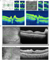
(A) Optical coherence tomography (OCT) (Cirrus, Zeiss) of the optic nerve in both eyes indicating mild infiltration of the optic nerve in both eyes. (B) OCT (Heidelberg Spectralis, Heidelberg) in the macula in both eyes indicating mild cystoid macular edema.
Figure 3.
Figure 3.
Congo red stain of fibroadipose tissue with small vessels showing congophilic amyloid deposition.

References

    1. Picken MM. Immunoglobulin light and heavy chain amyloidosis AL/AH: renal pathology and differential diagnosis. Contrib Nephrol. 2007;153:135-155. doi:10.1159/000096765 - DOI - PubMed
    1. Blancas-Mejía LM, Ramirez-Alvarado M. Systemic amyloidoses. Annu Rev Biochem. 2013;82:745-774. doi:10.1146/annurev-biochem-072611-130030 - DOI - PMC - PubMed
    1. Palladini G, Kyle RA, Larson DR, Therneau TM, Merlini G, Gertz MA. Multicentre versus single centre approach to rare diseases: the model of systemic light chain amyloidosis. Amyloid. 2005;12(2):120-126. doi:10.1080/13506120500107055 - DOI - PubMed
    1. Huang X, Wang Q, Jiang S, Chen W, Zeng C, Liu Z. The clinical features and outcomes of systemic AL amyloidosis: a cohort of 231 Chinese patients. Clin Kidney J. 2015;8(1):120-126. doi:10.1093/ckj/sfu117 - DOI - PMC - PubMed
    1. Booth DR, Stangou A, Williams RS, Gillmore JD, Tennent GA, Hawkins PN. Transthyretin Ile84Thr is associated with familial amyloid polyneuropathy. Hum Mutat. 2000;16(5):447. doi:10.1002/1098-1004(200011)16:5<447::AID-HUMU16>3.0.CO;2-P - DOI - PubMed

Publication types