

Stable and Reusable Lace-like Black Silicon Nanostructures Coated with Nanometer-Thick Gold Films for SERS-Based Sensing
- PMID: 37006910
- PMCID: PMC10043874
- DOI: 10.1021/acsanm.3c00281
Stable and Reusable Lace-like Black Silicon Nanostructures Coated with Nanometer-Thick Gold Films for SERS-Based Sensing
Abstract
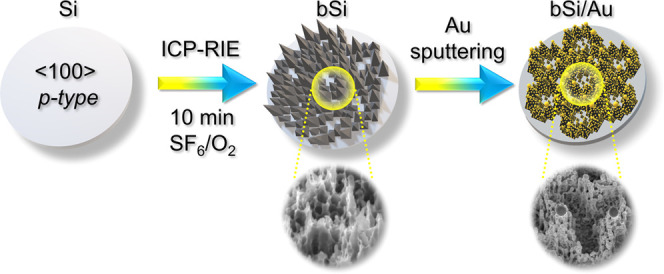
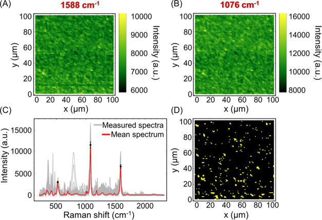
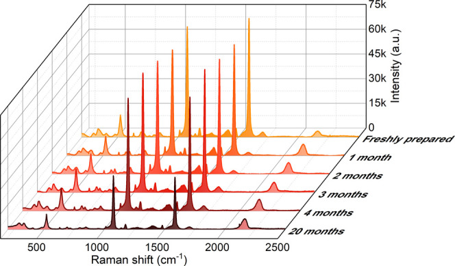
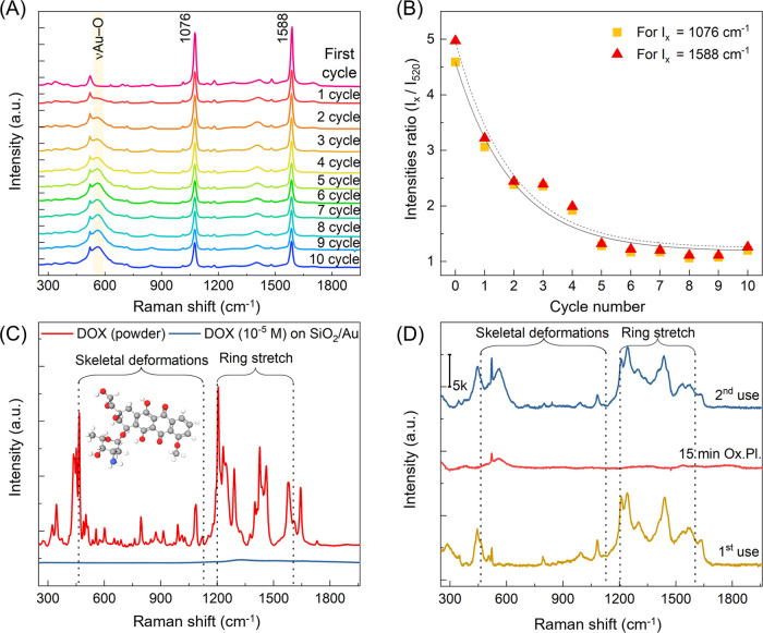
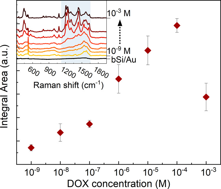
We propose a simple, fast, and low-cost method for producing Au-coated black Si-based SERS-active substrates with a proven enhancement factor of 106. Room temperature reactive ion etching of silicon wafer followed by nanometer-thin gold sputtering allows the formation of a highly developed lace-type Si surface covered with homogeneously distributed gold islands. The mosaic structure of deposited gold allows the use of Au-uncovered Si domains for Raman peak intensity normalization. The fabricated SERS substrates have prominent uniformity (with less than 6% SERS signal variations over large areas, 100 × 100 μm2). It has been found that the storage of SERS-active substrates in an ambient environment reduces the SERS signal by less than 3% in 1 month and not more than 40% in 20 months. We showed that Au-coated black Si-based SERS-active substrates can be reused after oxygen plasma cleaning and developed relevant protocols for removing covalently bonded and electrostatically attached molecules. Experiments revealed that the Raman signal of 4-MBA molecules covalently bonded to the Au coating measured after the 10th cycle was just 4 times lower than that observed for the virgin substrate. A case study of the reusability of the black Si-based substrate was conducted for the subsequent detection of 10-5 M doxorubicin, a widely used anticancer drug, after the reuse cycle. The obtained SERS spectra of doxorubicin were highly reproducible. We demonstrated that the fabricated substrate permits not only qualitative but also quantitative monitoring of analytes and is suitable for the determination of concentrations of doxorubicin in the range of 10-9-10-4 M. Reusable, stable, reliable, durable, low-cost Au-coated black Si-based SERS-active substrates are promising tools for routine laboratory research in different areas of science and healthcare.
© 2023 The Authors. Published by American Chemical Society.
Conflict of interest statement
The authors declare no competing financial interest.
Figures
Similar articles
-
Gold-capped silicon for ultrasensitive SERS-biosensing: Towards human biofluids analysis.Mater Sci Eng C Mater Biol Appl. 2018 Mar 1;84:208-217. doi: 10.1016/j.msec.2017.11.029. Epub 2017 Dec 5. Mater Sci Eng C Mater Biol Appl. 2018. PMID: 29519430
-
Trends in Application of SERS Substrates beyond Ag and Au, and Their Role in Bioanalysis.Biosensors (Basel). 2022 Nov 3;12(11):967. doi: 10.3390/bios12110967. Biosensors (Basel). 2022. PMID: 36354477 Free PMC article. Review.
-
SERS substrates fabricated with star-like gold nanoparticles for zeptomole detection of analytes.Nanoscale. 2015 Jun 14;7(22):10249-58. doi: 10.1039/c5nr02004b. Epub 2015 May 20. Nanoscale. 2015. PMID: 25990708
-
Gold-coated nanorod arrays as highly sensitive substrates for surface-enhanced raman spectroscopy.Langmuir. 2008 Dec 16;24(24):14172-5. doi: 10.1021/la802248t. Langmuir. 2008. PMID: 19053654
-
Advancements in reusable SERS substrates for trace analysis applications.Talanta. 2024 Nov 1;279:126640. doi: 10.1016/j.talanta.2024.126640. Epub 2024 Jul 31. Talanta. 2024. PMID: 39128272 Review.
Cited by
-
Black Silicon Surface-Enhanced Raman Spectroscopy Biosensors: Current Advances and Prospects.Biosensors (Basel). 2024 Sep 24;14(10):453. doi: 10.3390/bios14100453. Biosensors (Basel). 2024. PMID: 39451667 Free PMC article. Review.
References
-
- Kahraman M.; Mullen E. R.; Korkmaz A.; Wachsmann-Hogiu S. Fundamentals and Applications of SERS-Based Bioanalytical Sensing. Nanophotonics 2017, 6, 831–852. 10.1515/nanoph-2016-0174. - DOI
-
- Heeg S.; Mueller N. S.; Wasserroth S.; Kusch P.; Reich S. Experimental Tests of Surface-Enhanced Raman Scattering: Moving beyond the Electromagnetic Enhancement Theory. J. Raman Spectrosc. 2021, 52, 310–322. 10.1002/jrs.6014. - DOI
-
- He S.; Chua J.; Tan E. K. M.; Kah J. C. Y. Optimizing the SERS Enhancement of a Facile Gold Nanostar Immobilized Paper-Based SERS Substrate. RSC Adv. 2017, 7, 16264–16272. 10.1039/c6ra28450g. - DOI
LinkOut - more resources
Full Text Sources
Miscellaneous