

Diffuse large cell lymphoma of the lacrimal sac may mimic as acute dacryocystitis
- PMID: 37007254
- PMCID: PMC10062069
- DOI: 10.4103/ojo.ojo_304_21
Diffuse large cell lymphoma of the lacrimal sac may mimic as acute dacryocystitis
Abstract
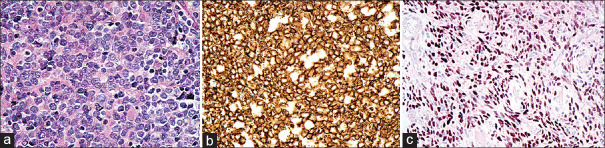
A 36-year-old male patient presented with a firm swelling in the left lacrimal sac region with a history of recurrent episodes of acute dacryocystitis, which partly resolved with systemic antibiotics. Computed tomography showed diffuse soft tissue mass without bony erosion in the same area. Incisional biopsy confirmed diffuse large cell lymphoma of non-Hodgkin's type by histopathology and immunohistochemistry. Oncologists did not detect systemic involvement, and the patient received six cycles of cyclophosphamide, hydroxydaunorubicin, oncovin, and prednisolone (CHOP). Epiphora was resolved, and no recurrence of the lesion was seen with subsequent dacryocystorhinostomy with intubation and was in good health for up to 3 years of follow-up. Although primary lacrimal sac lymphoma is a rare entity, high suspicion, and prompt action in atypical cases can save lives from aggressive diffuse large cell lymphoma.
Keywords: Acute dacryocystitis; CHOP therapy; dacryocystorhinostomy; diffuse large cell lymphoma.
Copyright: © 2023 Oman Ophthalmic Society.
Conflict of interest statement
There are no conflicts of interest.
Figures

References
-
- Yanoff M, Sassani J. Ocular Pathology. 8th. New York: Elsevier; 2018. pp. 232–3.
-
- Coupland SE, Hellmich M, Auw-Haedrich C, Lee WR, Stein H. Prognostic value of cell-cycle markers in ocular adnexal lymphoma: An assessment of 230 cases. Graefes Arch Clin Exp Ophthalmol. 2004;242:130–45. - PubMed
Publication types
LinkOut - more resources
Full Text Sources
Research Materials