Case Reports
. 2020 Nov 6;5(4):348-353.
doi: 10.1177/2474126420962649. eCollection 2021 Jul-Aug.

Pneumatic Vitreolysis With Intravitreal Air for Focal Vitreomacular Traction

Affiliations
Case Reports

Pneumatic Vitreolysis With Intravitreal Air for Focal Vitreomacular Traction

Mark E Seamone et al. J Vitreoretin Dis. .

Abstract

Purpose: To determine whether pneumatic vitreolysis with intravitreal air is effective for focal vitreomacular traction (VMT).

Methods: We conducted a retrospective consecutive case series of 20 eyes from 19 individuals with focal VMT who underwent pneumatic vitreolysis with intravitreal air (January 2017 to November 2018). We analyzed patients via spectral-domain optical coherence tomography before intravitreal air injection and at 1 month. The primary outcome measure was release of VMT.

Results: We observed release of VMT in 55% of individuals. An analysis limited to phakic eyes demonstrated release of VMT in 69%, and 65% developed improved best-corrected visual acuity. Individuals with persistent VMT and visual improvement had a significant reduction in angle of vitreoretinal insertion (P < .01), area under VMT (P < .05), and subfoveal cyst area (P < .05).

Conclusions: Intravitreal air is an effective treatment for focal VMT. In individuals with persistent VMT, visual-acuity improvement was associated with a reduction in overall VMT.

Keywords: intravitreal air; pneumatic vitreolysis; vitreomacular traction.


Conflict of interest statement

The author(s) declare no conflict of interest with respect to the research, authorship, and/or publication of this article.

Figures
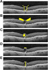
Figure 1.
Figure 1.
Analysis of the vitreoretinal interface. (A) Angle of vitreous insertion was measured using a line perpendicular to the retinal pigment epithelium. (B) Area under vitreomacular traction measured 750 µm on either side of the foveal center. (C) Subfoveal cyst area. (D) Horizontal length of vitreomacular adhesion. (E) Retinal thickness defined by the boundaries of the internal limiting membrane and the retinal pigment epithelium.
Figure 2.
Figure 2.
Intravitreal air injection causes a decrease in the angle of vitreous insertion in individuals with persistent vitreomacular traction and visual improvement. (A) Representative example of the average angle of vitreous insertion in a single individual prior to intravitreal air injection. (B) Average angle of vitreous insertion for the same individual at 1 month following intravitreal air injection.
Figure 3.
Figure 3.
The angle of vitreous insertion is decreased in individuals with persistent vitreomacular traction (VMT) and visual improvement following intravitreal air injection. The angle of vitreous insertion was measured relative to a vertical line perpendicular to the retinal pigment epithelium. Measurements were taken using spectral-domain optical coherence tomography prior to intravitreal air injection and at 1 month following injection. (A) Individuals with persistent VMT and visual improvement (n = 4, **P < .01; 95% CI, 4.5-12.6). (B) Individuals with persistent VMT and no visual improvement (n = 3, not significant [NS] = P > .05; 95% CI, –23.4 to 18.6).
Figure 4.
Figure 4.
Area under vitreomacular traction (VMT) is decreased in individuals with persistent VMT and visual improvement following intravitreal air injection. The area between the posterior hyaloid face and the internal limiting membrane was measured within 750 µm on either side of the foveal center. Measurements were taken using spectral-domain optical coherence tomography prior to intravitreal air injection and at 1 month following injection. (A) Individuals with persistent VMT and visual improvement (n = 4, *P < .05; 95% CI, 9.1-40.9). (B) Individuals with persistent VMT and no visual improvement (n = 3, not significant [NS] = P > .05; 95% CI, –164.1 to 90.8).
Figure 5.
Figure 5.
Subfoveal cyst area is decreased in individuals with persistent vitreomacular traction (VMT) and visual improvement following intravitreal air injection. The area of the subfoveal cystic space was measured using spectral-domain optical coherence tomography prior to intravitreal air injection and at 1 month following injection. (A) Individuals with persistent VMT and visual improvement (n = 4, *P < .05; 95% CI, 2.3-32.0). (B) Individuals with persistent VMT and no visual improvement (n = 3, not significant [NS] = P > .05; 95% CI, –101.4 to 161.4).

References

    1. Folk JC, Adelman RA, Flaxel CJ, Hyman L, Pulido JS, Olsen TW. Idiopathic epiretinal membrane and vitreomacular traction Preferred Practice Pattern® guidelines. Ophthalmology. 2016;123(1):P152–P181. doi:10.1016/j.ophtha.2015.10.048 - PubMed
    1. Duker JS, Kaiser PK, Binder S, et al. The International Vitreomacular Traction Study Group classification of vitreomacular adhesion, traction, and macular hole. Ophthalmology. 2013;120(12):2611–2619. doi:10.1016/j.ophtha.2013.07.042 - PubMed
    1. Tzu JH, John VJ, Flynn HW, et al. Clinical course of vitreomacular traction managed initially by observation. Ophthalmic Surg Lasers Imaging Retin. 2015;46(5):571–576. doi:10.3928/23258160-20150521-09 - PubMed
    1. Patron ME, Baumal CR, Rogers AH, et al. Anatomic and visual outcomes of vitrectomy for vitreomacular traction syndrome. Ophthalmic Surg Lasers Imaging. 2010;41(4):425–431. doi:10.3928/15428877-20100525-07 - PubMed
    1. Dugel PU, Tolentino M, Feiner L, Kozma P, Leroy A. Results of the 2-Year Ocriplasmin for Treatment for Symptomatic Vitreomacular Adhesion Including Macular Hole (OASIS) randomized trial. Ophthalmology. 2016;123(10):2232–2247. doi:10.1016/j.ophtha.2016.06.043 - PubMed

Publication types