. 2023 Mar 20:10:102142.
doi: 10.1016/j.mex.2023.102142. eCollection 2023.

A thermally robust method of sample sealing for capillary DLS

Affiliations

A thermally robust method of sample sealing for capillary DLS

Sharla Harvey et al. MethodsX. .

Abstract

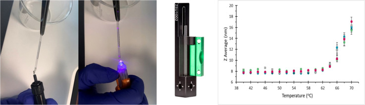
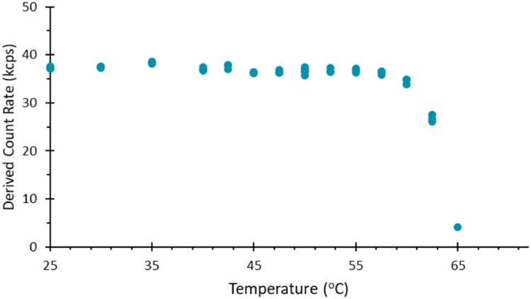
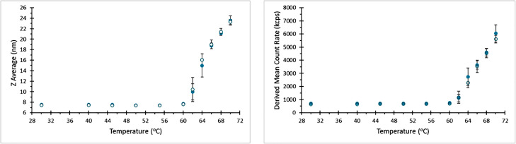
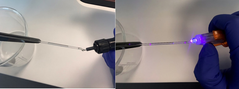
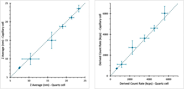
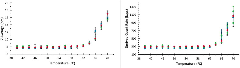
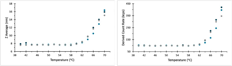
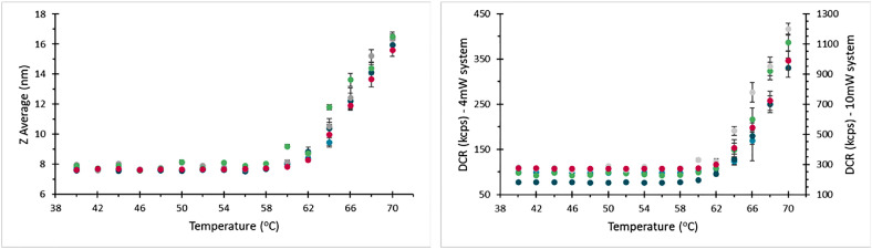
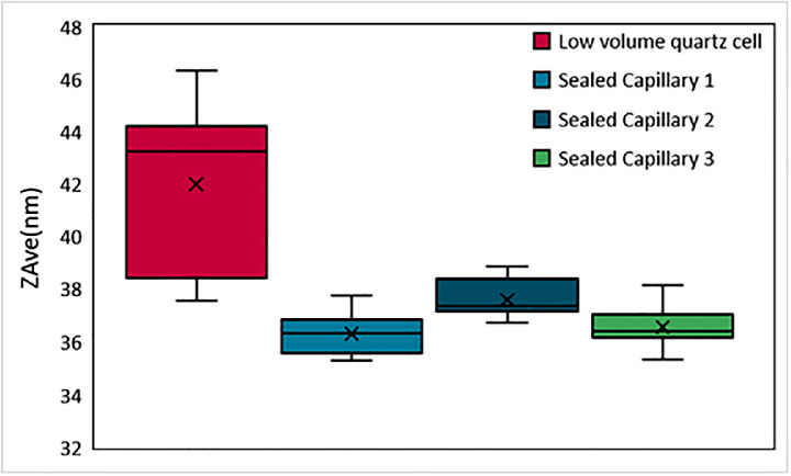
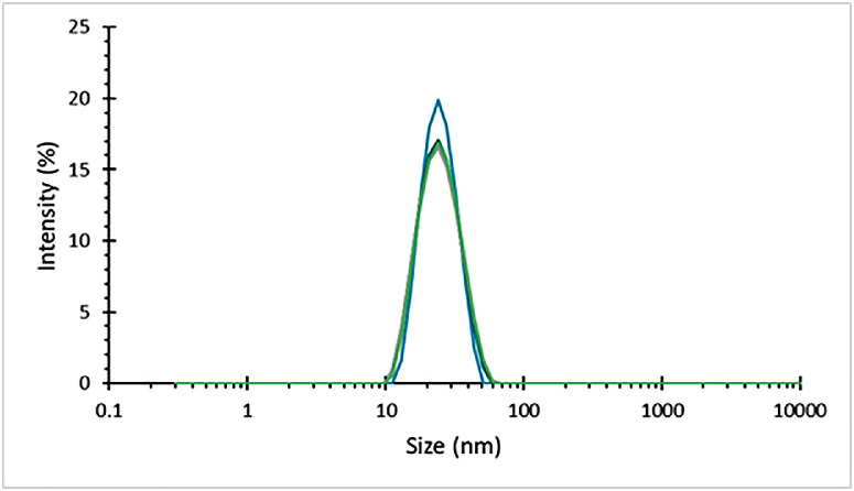
Capillary Dynamic Light Scattering (DLS), has recently been introduced as a simple and enabling technique that increases the measurement range of traditional DLS analysis with minimized sample volumes (Ruseva et al., 2018). The previously published protocol for the preparation of samples for analysis within a capillary called for sealing of the capillary end using a clay compound (Ruseva et al., 2019). This material is not, however, compatible with organic solvents, nor with elevated sample temperatures. To extend the uses of capillary DLS to more complex assays like thermal aggregation studies, a new sealing method is demonstrated using a UV curing compound. This further motivates the use of capillary DLS to minimize volumes of destroyed precious samples in pharmaceutical development assays to study thermal kinetics.•Use of UV curing compound to seal capillaries used in DLS to preserve low volumes of sample.

Keywords: An optimized method of sample sealing for capillary Dynamic Light Scattering; Capillary; Dynamic light scattering; Thermal ramps.


Conflict of interest statement

The authors declare the following financial interests/personal relationships which may be considered as potential competing interests. The authors are employees of Malvern Panalytical Ltd. but declare no other competing interests.

Figures

Image, graphical abstract
Graphical abstract
Fig 1
Fig. 1
Derived mean count rate as a function of sample temperature for a 1 mg/ml dispersion of BSA, recorded for a capillary sealed with clay. The onset of fluctuations in the detected scattering demonstrates the breakdown of the clay compound, most notably after 40 °C. During this experiment, the intended end temperature was 70 °C. However, above 60 °C, the sample was lost, and no further data was recorded.
Fig 2
Fig. 2
Intensity weighted particle size distribution for a 1 mg/ml dispersion of BSA, recorded in several capillaries sealed using wax. All these measurements were performed at 25 °C such that no sample aggregation was expected. However, shifts in peak size and presence of larger material is evident, suggesting that the heat used to melt the wax during sealing has influenced the sample.
Fig 3
Fig. 3
Intensity weighted particle size distribution for a 1 mg/ml dispersion of BSA, recorded in several capillaries sealed using UV curing resin, showing good quality repeatable data, with no apparent effect on the sample from either the resin or the UV light during sealing.
Fig 4
Fig. 4
Derived mean count rate as a function of temperature for a BSA sample during a temperature ramp applied to a capillary where one end is sealed using UV curing compound. At elevated temperatures the scattering intensity.
Fig 5
Fig. 5
Comparison of thermograms for BSA recorded using a capillary sealed at both ends using UV curing resin (Filled symbols), and a low volume quartz cell (Hollow symbols).
Fig 6
Fig. 6
Left: Small amount of resin being administered onto the tip of the capillary. Right: UV light being used to set the resin which takes approximately 5 s.
Fig 7
Fig. 7
Comparison of reported size and derived count rate for thermal ramps of a BSA sample, recorded using a sealed capillary and a low volume quartz cell. Note that error bars increase significantly in scale during aggregation of the sample due to sample variability during this process.
Fig 8
Fig. 8
Thermograms for repeat measurements on a given instrument and user.
Fig 9
Fig. 9
Thermograms recorded for samples loaded by separate operators, demonstrating good reproducibility.
Fig 10
Fig. 10
Thermograms recorded on different instruments, showing agreement between these measurements. Note the magenta data set here is recorded on a system fitted with a 10 mW laser, while the remainder of records here were for 4 mW systems, so the absolute light scattering between these systems is different, but the same trend is followed in each case.
Fig 11
Fig. 11
Deviation from an average thermogram for all temperature trend measurements for BSA, demonstrating variation based on instrument, user and capillary.
Fig 12
Fig. 12
Measured average size of a silica sample, in a quartz cell and three separate sealed capillaries, with size measurements recorded at temperatures ranging from 25 to 90 °C.
Fig 13
Fig. 13
Intensity weighted particle size distribution for 200 kDa polystyrene dispersed in toluene, in a sealed capillary, measured approximately 24 h after preparation of the capillary.

References

    1. Ruseva V., et al. Capillary dynamic light scattering: continuous hydrodynamic particle size from the nano to the micro-scale. Colloids Surf. A. 2018;558:504–511.
    1. Ruseva V., Jankevics H., Corbett J. An optimized filling method for capillary DLS. MethodsX. 2019;6:p606–p614. - PMC - PubMed
    1. Markova N., et al. Biophysical characterization of viral and lipid-based vectors for vaccines and therapeutics with light scattering and calorimetric techniques. Vaccines. 2022;10(1):49. - PMC - PubMed
    1. Dauer K., et al. Microwell plate-based dynamic light scattering as a high-throughput characterization tool in biopharmaceutical development. Pharmaceutics. 2021;13:172. - PMC - PubMed
    1. Malm A., Corbett &.J. Improved dynamic light scattering using an adaptive and statistically driven time resolved treatment of correlation data. Sci. Rep. 2019;9:13519. - PMC - PubMed

LinkOut - more resources