. 2022 Nov 29:61:e61.
doi: 10.6620/ZS.2022.61-61. eCollection 2022.

Expressions of the Protein Phosphatases PP1 and PP4 during the Embryonic Diapause Process of the Silkworm Bombyx mori

Affiliations

Expressions of the Protein Phosphatases PP1 and PP4 during the Embryonic Diapause Process of the Silkworm Bombyx mori

Shi-Hong Gu et al. Zool Stud. .

Abstract

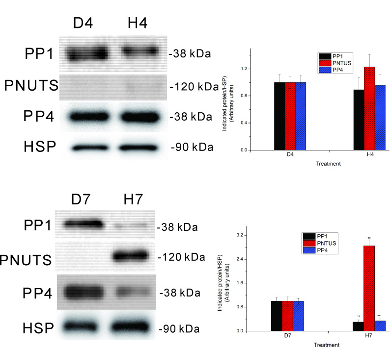
Reversible protein phosphorylation is accomplished by the opposing activities of kinases and phosphatases. We previously demonstrated the regulation of serine/threonine protein phosphatase (PP) type 2A (PP2A) and 2B (PP2B or calcineurin) during the embryonic diapause process of Bombyx mori. In the present study, we further examine the expressions of other PPs (PP1 and PP4) during embryonic stages. An immunoblot analysis showed that Bombyx eggs contained a 38-kDa PP1 catalytic subunit (PP1-C), a 38-kDa PP4 catalytic subunit (PP4-C), and a 120-kDa PP1 nuclear targeting subunit (PNUTS), each of which underwent differential changes between diapause and developing eggs during the embryonic process. In non-diapause eggs, eggs whose diapause initiation was prevented by HCl, and eggs in which diapause had been terminated by chilling diapausing eggs at 5°C for 70 days and then were transferred to 25°C, protein levels of PP1-C and PP4-C remained relatively high during the early embryonic stages and then decreased during middle (for PP1-C) or later (for PP4-C) embryonic stages. However, protein levels of PP1-C and PP4-C in diapause eggs remained at high levels during the first 8 days after oviposition. PNUTS protein levels showed inverse temporal changes, with increased levels being detected during the later embryonic stages of developing eggs. The direct determination of PP1 enzymatic activity showed higher activity in developing eggs than in diapause eggs. Examination of temporal changes in mRNA expression levels of PP1-C and PP4-C showed no difference between HCl-treated and diapause eggs. These results indicated that differential protein levels of PP1-C/PNUTS and PP4-C, and increased enzymatic activity of PP1 were likely related to the embryonic development of B. mori.

Keywords: Bombyx mori; Diapause; Gene expression; PNUTS; PP1; PP4; Phosphatase.


Figures

Fig. 1.
Fig. 1.
Western blot analysis of PP1-C, PNUTS, and PP4-C in silkworm eggs. Following SDS-PAGE and immunoblotting, silkworm egg extracts (a quarter of one egg for each lane) were probed with anti-PP1α (PP1), anti-PNUTS (PNUTS), anti-PP4-C (PP4), and anti-HSP 90 (HSP) antibodies. (A) Results from day 4. D4, extracts from day 4 diapause eggs; H4, extracts from eggs whose diapause was prevented by HCl (4 days after HCl treatment). (B) Results from day 7. D7, extracts from day 7 diapause eggs; H7, extracts from eggs whose diapause was prevented by HCl (7 days after HCl treatment). Results shown in the left panel are representative of four independent experiments. Each protein band was quantified and normalized to the level of HSP in the right panel. Asterisks indicate significant differences compared to respective diapause eggs (by Student’s t-test, ** p < 0.01).
Fig. 2.
Fig. 2.
Effect of HCl treatment on protein levels of PP1-C, PNUTS, and PP4-C during the first 8 days after treatment. Diapause-destined eggs that had been incubated at 25°C for 20 h after oviposition were treated with HCl and then incubated at 25°C. Egg lysates from each stage were prepared and subjected to an immunoblot analysis with anti-PP1α (PP1), anti-PNUTS (PNUTS), anti-PP4-C (PP4), and HSP 90 (HSP) antibodies. Lysates from a quarter of one egg were used for each lane. Molecular weight markers are shown on the right side of the gel (A). Results shown in the upper panel are representative of four independent experiments. Quantified protein levels relative to HSP were standardized to levels from the first day after HCl treatment. Different letters above the bars indicate significant differences (ANOVA followed by Tukey’s test).
Fig. 3.
Fig. 3.
Western blot analysis of protein levels of PP1-C, PNUTS, and PP4-C in diapause eggs. Egg lysates from each stage after oviposition were prepared and subjected to an immunoblot analysis with anti-PP1α (PP1), anti-PNUTS (PNUTS), anti-PP4-C (PP4), and anti-HSP 90 (HSP) antibodies. Lysates from a quarter of one egg were used for each lane. Molecular weight markers are shown on the right side of the gel (A). Results shown in the upper panel are representative of four independent experiments. Quantified protein levels relative to HSP were standardized to levels from the first day after oviposition. Different letters above the bars indicate significant differences (ANOVA followed by Tukey’s test).
Fig. 4.
Fig. 4.
Western blot analysis of protein levels of PP1-C, PNUTS, and PP4-C in non-diapause eggs. Egg lysates from each stage after oviposition were prepared and subjected to an immunoblot analysis with anti-PP1α (PP1), anti-PNUTS (PNUTS), anti-PP4-C (PP4), and anti-HSP 90 (HSP) antibodies. Lysates from a quarter of one egg were used for each lane. Molecular weight markers are shown on the right side of the gel (A). Results shown in the upper panel are representative of four independent experiments. Quantified protein levels relative to HSP were standardized to levels from the first day after oviposition. Different letters above the bars indicate significant differences (ANOVA followed by Tukey’s test).
Fig. 5.
Fig. 5.
Western blot analysis of protein levels of PP1-C, PNUTS, and PP4-C in eggs in which diapause had been terminated by chilling of diapausing eggs at 5°C for 70 days and then transferred to 25°C. Egg lysates from each stage after being transferred to 25°C were prepared and subjected to an immunoblot analysis with anti-PP1α (PP1), anti-PNUTS (PNUTS), anti-PP4-C (PP4), and anti-HSP 90 (HSP) antibodies. Lysates from a quarter of one egg were used for each lane. Molecular weight markers are shown on the right side of the gel (A). Results shown in the upper panel are representative of four independent experiments. Quantified protein levels relative to HSP were standardized to levels from the first day after transfer to 25°C. Different letters above the bars indicate significant differences (ANOVA followed by Tukey’s test).
Fig. 6.
Fig. 6.
Changes in PP1 enzymatic activity in Bombyx eggs. A, diapause eggs; B, non-diapause eggs; C, eggs whose diapause initiation was prevented by HCl; D, eggs in which diapause had been terminated by chilling. Egg extracts from each stage were prepared, and PP1 enzymatic activity was determined with a PP1 assay kit. The data represent mean ± SEM of four separate assays.
Fig. 7.
Fig. 7.
Changes in mRNA expression levels of PP1-C (A) and PP4-C (B) in diapause eggs and diapause-destined eggs treated with HCl 20 h after oviposition to prevent the eggs from entering diapause. Egg extracts from each stage were then prepared, and mRNA expression levels were determined by qRT-PCR. The mRNA expression levels of PP1-C and PP4-C relative to rp49 were standardized to the means of day 0 of diapause eggs and are represented as the relative amount ± SEM (n = 4). Circles, HCl-treated eggs; squares, diapause eggs.
Fig. 8.
Fig. 8.
Our current understanding of the signaling network involved in the Bombyx embryonic diapause process. See text for details.

Similar articles

References

    1. Allen PB, Kwon YG, Nairn AC, Greengard P. 1998. Isolation and characterization of PNUTS, a putative protein phosphatase 1 nuclear targeting subunit. J Biol Chem 273:4089–4095. doi:10.1074/jbc.273.7.4089. - PubMed
    1. Axton JM, Dombrádi V, Cohen PT, Glover DM. 1990. One of the protein phosphatase 1 isoenzymes in Drosophila is essential for mitosis. Cell 63:33–46. doi:10.1016/0092-8674(90)90286-n. - PubMed
    1. Baker SH, Frederick DL, Bloecher A, Tatchell K. 1997. Alanine scanning mutagenesis of protein phosphatase type 1 in the yeast Saccharomyces cerevisiae. Genetics 145:615–626. doi:10.1093/genetics/145.3.615. - PMC - PubMed
    1. Bhaskar PT, Hay N. 2007. The two TORCs and Akt. Dev Cell 12:487–502. doi:10.1016/j.devcel.2007.03.020. - PubMed
    1. Bollen M, Peti W, Ragusa MJ, Beullens M. 2010. The extended PP1 toolkit: designed to create specificity. Trends Biochem Sci 35:450–458. doi:10.1016/j.tibs.2010.03.002. - PMC - PubMed

LinkOut - more resources