. 2023 Mar 10:14:1073138.
doi: 10.3389/fgene.2023.1073138. eCollection 2023.

A comprehensive pan-cancer analysis unveiling the oncogenic effect of plant homeodomain finger protein 14 (PHF14) in human tumors

Affiliations

A comprehensive pan-cancer analysis unveiling the oncogenic effect of plant homeodomain finger protein 14 (PHF14) in human tumors

Zhiyou Cao et al. Front Genet. .

Abstract

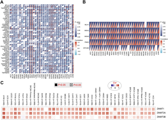
The plant homeodomain (PHD) finger refers to a protein motif that plays a key role in the recognition and translation of histone modification marks by promoting gene transcriptional activation and silencing. As an important member of the PHD family, the plant homeodomain finger protein 14 (PHF14) affects the biological behavior of cells as a regulatory factor. Several emerging studies have demonstrated that PHF14 expression is closely associated with the development of some cancers, but there is still no feasible pan-cancer analysis. Based on existing datasets from the Cancer Genome Atlas (TCGA) and the Gene Expression Omnibus (GEO), we performed a systematic analysis of the oncogenic role of the PHF14 gene in 33 human cancers. The expression level of PHF14 was significantly different between different types of tumors and adjacent normal tissues, and the expression or genetic alteration of PHF14 gene was closely related to the prognosis of most cancer patients. Levels of cancer-associated fibroblasts (CAFs) infiltration in various cancer types were also observed to correlate with PHF14 expression. In some tumors, PFH14 may play a role in tumor immunity by regulating the expression levels of immune checkpoint genes. In addition, the results of enrichment analysis showed that the main biological activities of PHF14 were related to various signaling pathways or chromatin complex effects. In conclusion, our pan-cancer research shows that the expression level of PHF14 is closely related to the carcinogenesis and prognosis of certain tumors, which needs to be further verified by more experiments and more in-depth mechanism exploration.

Keywords: PHF14; cancer; carcinogenesis; gene expression; pan-cancer analysis; prognosis.


Conflict of interest statement

The authors declare that the research was conducted in the absence of any commercial or financial relationships that could be construed as a potential conflict of interest.

Figures

FIGURE 1
FIGURE 1
The PHF14 expression status in different tumors and normal tissues. (A) Consensus PHF14 tissue expression based on datasets of HPA (Human Protein Atlas), GTEx, and FANTOM5 (function annotation of the mammalian genome). (B) Consensus PHF14 cell type expression based on the above datasets. (C,D) The TCGA project’s PHF14 gene expression difference in different tumors or specific tumor subtype tissues and unpaired or paired adjacent normal tissues was analyzed by TIMER2. *p < 0.05; **p < 0.01; ***p < 0.001. (E) Difference of the PHF14 total protein expression between normal and tumor tissues of breast cancer, colon cancer, LUAD, clear cell RCC, UCEC, GBM, HNSC, LIHC, PAAD and ovarian cancer were analyzed based on the CPTAC dataset. *p < 0.05; ***p < 0.001.
FIGURE 2
FIGURE 2
On the basis of the TCGA dataset, (A–F) analysis of the expression level of PHF14 gene in the different pathological stages (stage I, II, III, and IV) in BRCA, COAD, KICH, SKCM, LIHC and STAD tumors by applying GEPIA2.
FIGURE 3
FIGURE 3
Correlation between PHF14 gene expression and survival prognosis for tumors in TCGA analyzed with the GEPIA2 tool. (A) Overall survival analysis. (B) Disease-free survival. (C–F) Progression-free survival in KIRC, LGG, LUAD, and PAAD. The results with significant differences were visualized through a survival map and Kaplan-Meier curves.
FIGURE 4
FIGURE 4
Mutation characteristics of PHF14 gene in different kind of tumors of TCGA were analyzed by the cBioPortal tool. (A) The mutation type and alteration frequency in various cancers. (B) The mutation sites in PHF14. (C–H) The correlation between MSH6 mutation status and overall, disease-specific, and progression-free survival prognoses of BRCA and CESC.
FIGURE 5
FIGURE 5
Correlation analysis between PHF14 expression and immune infiltration of cancer-associated fibroblasts. (A) The correlation between PHF14 expression and immune infiltration of cancer-associated fibroblasts for all TCGA tumors evaluated by different algorithms, including EPIC, MCPCOUNTER and TIDE. (B) Scatter plot data for selected tumors generated using one of the algorithms were provided.
FIGURE 6
FIGURE 6
Correlation analysis of PHF14 expression and immune checkpoint genes in pan-cancer. (A) Correlation analysis between PHF14 expression in Pan-cancer and immune checkpoint gene expression. (B) Correlation analysis of PFH14 expression and the expression levels of five common MMRs genes (MLH1, MSH2, MSH6, PMS2, EPCAM) in various types of tumors in TCGA. (C) Corresponding heatmap data for targeted genes (DNMT1, DNMT3A, DNMT3B) in selected cancer types in TCGA.
FIGURE 7
FIGURE 7
PHF14-related gene function enrichment analysis. (A) On the basis of the STRING tool, Co-expression network of 50 genes co-expressed with PHF14 were obtained. (B) CC, BP, MF and KEGG pathways analysis based on PHF14-correlated genes and PHF14-binding protein. (C–E) Results of GSEA of the top 3 rankings of PHF14 correlation with signaling pathways in KEGG database.
FIGURE 8
FIGURE 8
Validation of PHF14 gene at translational and transcriptional levels. (A–C) Immunohistochemistry images from The Human Protein Atlas database were used to verify the translation expression level of PHF14 gene in KIRC, PAAD, and LIHC. The figures showed that PHF14 protein was highly expressed in the above three kinds of tumor tissues compared with the corresponding normal tissues. (D–F) Real-time PCR analysis of PHF14 transcription levels in KIRC, PAAD and LIHC showed that the mRNA expression level of PHF14 in human kidney renal clear cell carcinoma cell lines (Caki-2 and 786-O) was higher than that in human renal tubular epithelial cell line (HK-2). And the mRNA expression of PHF14 in human pancreatic adenocarcinoma cell lines (ASPC-1 and PANC-1) and human liver hepatocellular carcinoma cell lines (MHCC-97h and Huh-7) was higher than that of human pancreatic duct epithelial cell line (hTERT-HPNE) and human normal liver cell line (LO2), respectively. **p < 0.01; ***p < 0.001; ****p < 0.0001.
FIGURE 9
FIGURE 9
PHF14-knockdown inhibited the growth, migration and invasion of PANC-1 and MHCC-97h cells, and promoted cells apoptosis. (A) The knockdown efficacy of PHF14 examined in PANC-1 and MHCC-97h cells by qRT-PCR. (B) The effect of the siRNA targeting of PHF14 on cell proliferation was measured with the MTT assay at the indicated times following transfection. (C, D) Ten thousand PANC-1 and MHCC-97h cells per well were seeded in 96-well plates overnight. EdU and Hoechest co-staining was performed on the second day to assess cellular DNA replication activities (Scale bar = 200 μm). (E) Values are counted as EdU+ cells/total cells. (F, G) Cell suspensions (the cell density was adjusted to 1-10*105/mL) of 200 μL PANC-1 and MHCC-97h were seeded into the upper chamber of a transwell insert (Scale bar = 100 μm). After 24 h of incubation, cells which had adhered to the lower membrane of the inserts were fixed, stained with 1% crystal violet and counted for analysis. (H) Flow cytometry was used to detect cell apoptosis in PHF14- and negative control-knockdown cells through annexin V-FITC/PI staining. (I) Wound closure scratch was used to assess the effect of PHF14 on the migratory capacity of PANC-1 and MHCC-97h cells. Each assay was performed in triplicate (**p < 0.01; ***p < 0.001; ****p < 0.0001).

Similar articles

Cited by

References

    1. Aasland R., Gibson T. J., Stewart A. F. (1995). The PHD finger: Implications for chromatin-mediated transcriptional regulation. Trends Biochem. Sci. 20 (2), 56–59. 10.1016/s0968-0004(00)88957-4 - DOI - PubMed
    1. Akazawa T., Yasui K., Gen Y., Yamada N., Tomie A., Dohi O., et al. (2013). Aberrant expression of the PHF14 gene in biliary tract cancer cells. Oncol. Lett. 5 (6), 1849–1853. 10.3892/ol.2013.1278 - DOI - PMC - PubMed
    1. Asplund A., Edqvist P. H. D., Schwenk J. M., Ponten F. (2012). Antibodies for profiling the human proteome-The Human Protein Atlas as a resource for cancer research. Proteomics 12 (13), 2067–2077. 10.1002/pmic.201100504 - DOI - PubMed
    1. Ballare C., Lange M., Lapinaite A., Martin G. M., Morey L., Pascual G., et al. (2012). Phf19 links methylated Lys36 of histone H3 to regulation of Polycomb activity. Nat. Struct. Mol. Biol. 19 (12), 1257–1265. 10.1038/nsmb.2434 - DOI - PMC - PubMed
    1. Barrett T., Wilhite S. E., Ledoux P., Evangelista C., Kim I. F., Tomashevsky M., et al. (2013). NCBI GEO: Archive for functional genomics data sets--update. Nucleic Acids Res. 41, D991–D995. (Database issue. 10.1093/nar/gks1193 - DOI - PMC - PubMed