. 2023 Mar 16:11:1152207.
doi: 10.3389/fbioe.2023.1152207. eCollection 2023.

Mesenchymal stem cell-derived secretome enhances nucleus pulposus cell metabolism and modulates extracellular matrix gene expression in vitro

Affiliations

Mesenchymal stem cell-derived secretome enhances nucleus pulposus cell metabolism and modulates extracellular matrix gene expression in vitro

Veronica Tilotta et al. Front Bioeng Biotechnol. .

Abstract

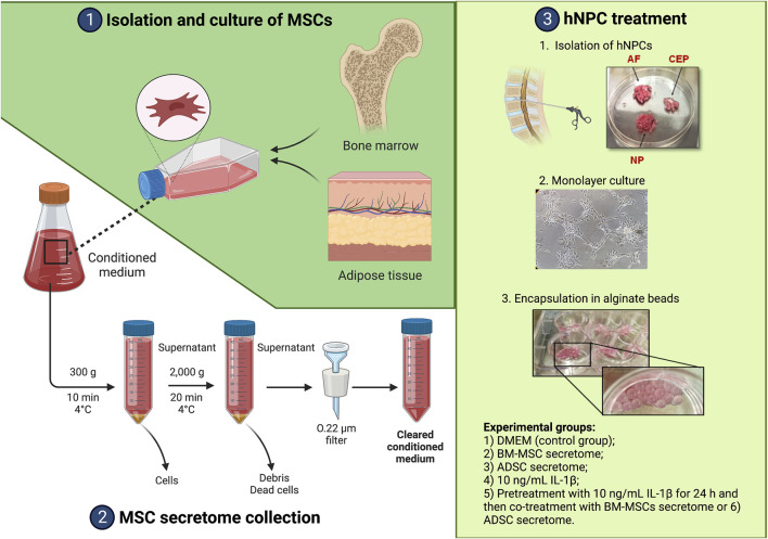
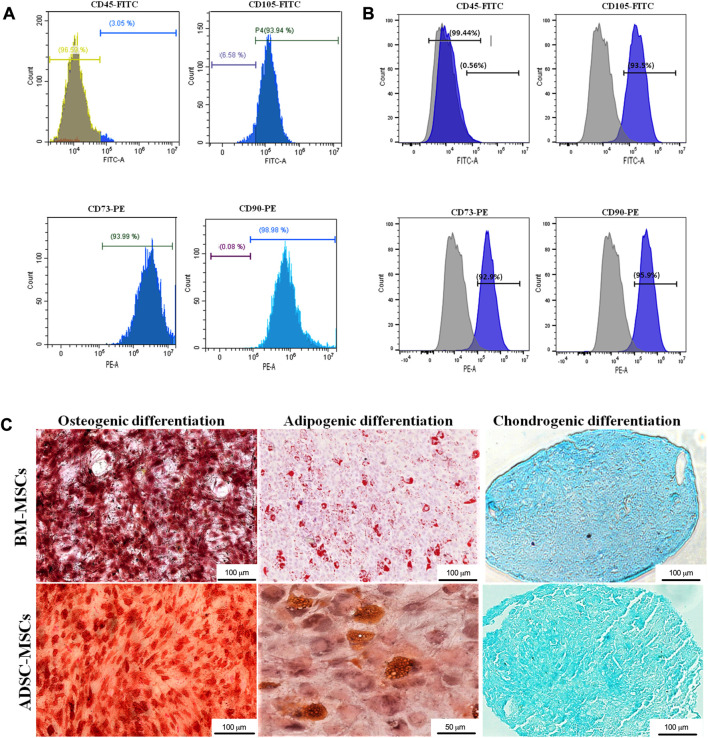
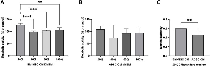
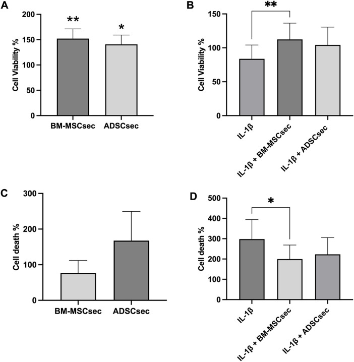
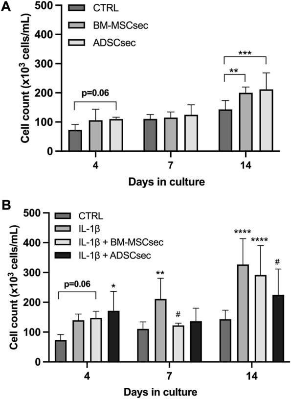
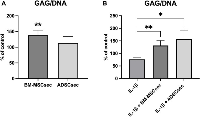
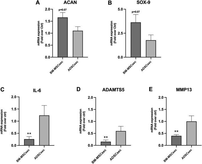
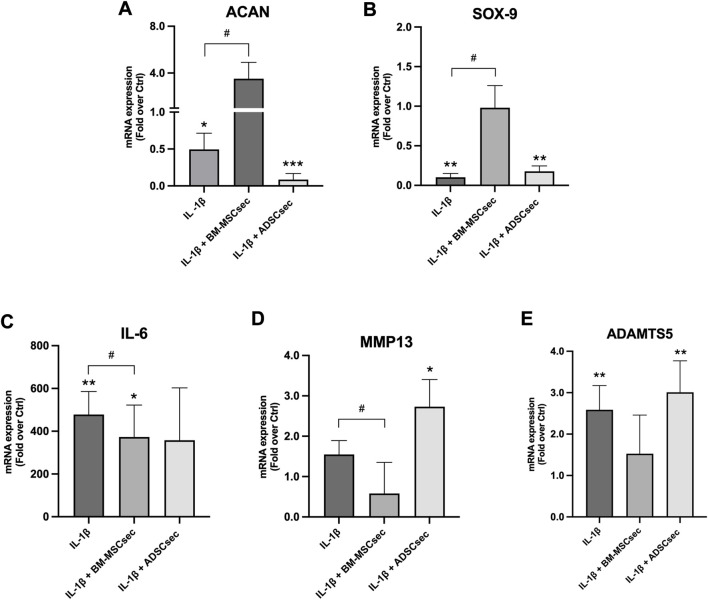
Introduction: Intradiscal mesenchymal stromal cell (MSC) therapies for intervertebral disc degeneration (IDD) have been gaining increasing interest due to their capacity to ameliorate intervertebral disc metabolism and relieve low back pain (LBP). Recently, novel investigations have demonstrated that most of MSC anabolic effects are exerted by secreted growth factors, cytokines, and extracellular vesicles, collectively defined as their secretome. In this study, we aimed to evaluate the effect of bone-marrow-MSCs (BM-MSCs) and adipose-derived stromal cells (ADSCs) secretomes on human nucleus pulposus cells (hNPCs) in vitro. Methods: BM-MSCs and ADSCs were characterized according to surface marker expression by flow cytometry and multilineage differentiation by Alizarin red, Red Oil O and Alcian blue staining. After isolation, hNPCs were treated with either BM-MSC secretome, ADSC secretome, interleukin (IL)-1β followed by BM-MSC secretome or IL-1β followed by ADSC secretome. Cell metabolic activity (MTT assay), cell viability (LIVE/DEAD assay), cell content, glycosaminoglycan production (1,9-dimethylmethylene blue assay), extracellular matrix and catabolic marker gene expression (qPCR) were assessed. Results: 20% BM-MSC and ADSC secretomes (diluted to normal media) showed to exert the highest effect towards cell metabolism and were then used in further experiments. Both BM-MSC and ADSC secretomes improved hNPC viability, increased cell content and enhanced glycosaminoglycan production in basal conditions as well as after IL-1β pretreatment. BM-MSC secretome significantly increased ACAN and SOX9 gene expression, while reducing the levels of IL6, MMP13 and ADAMTS5 both in basal conditions and after in vitro inflammation with IL-1β. Interestingly, under IL-1β stimulation, ADSC secretome showed a catabolic effect with decreased extracellular matrix markers and increased levels of pro-inflammatory mediators. Discussion: Collectively, our results provide new insights on the biological effect of MSC-derived secretomes on hNPCs, with intriguing implications on the development of cell-free approaches to treat IDD.

Keywords: growth factors; intervertebral disc; intervertebral disc degeneration; low back pain; mesenchymal stromal cells; secretome.


Conflict of interest statement

The authors declare that the research was conducted in the absence of any commercial or financial relationships that could be construed as a potential conflict of interest.

Figures

FIGURE 1
FIGURE 1
Summary of the experimental workflow. Created with www.biorender.com.
FIGURE 2
FIGURE 2
Characterization of MSCs. BM-MSCs (A) and ADSCs (B) where characterized with flow cytometry through immunophenotype analysis of MSC cell surface markers CD45, CD105, CD73, CD90. (C) Osteogenic, adipogenic, and chondrogenic differentiation was determined by Alizarin red, Oil red O and Alcian blue staining, respectively (magnification: ×20, scale bars: 50 μm and 100 μm).
FIGURE 3
FIGURE 3
BM-MSC-derived secretome enhanced hNPC metabolic activity. MTT assay on hNPCs treated with BM-MSC-derived CM (A) or ADSC-derived CM (B) at a 20%, 40%, 80% or 100% concentration (percentage of CM diluted in DMEM, volume/volume) showed that 20% BM-MSC CM yielded the greatest effect towards cell metabolic activity. Data are expressed as percent variation between the control and experimental groups. (C) 20% BM-MSC-derived CM determined a significantly higher increase of metabolic activity in hNPCs compared to 20% ADSC-CM. **p < 0.01, ***p < 0.001, ****p < 0.0001.
FIGURE 4
FIGURE 4
MSC-derived secretome enhanced hNPC viability and reduced cell death. The LIVE/DEAD assay showed that both BM-MSCsec and ADSCsec significantly increased cell viability compared to the control group (A). Following IL-1β pretreatment, BM-MSCsec significantly rescued hNPC vitality compared to cells treated with IL-1β alone (B). (C) BM-MSCsec and ADSCsec treatment did not substantially impact on cell death. (D) After IL-1β pretreatment, BM-MSCsec significantly reduced the number of dead cells compared to hNPCs treated with IL-1β alone. Results were normalized to the control group and expressed as percent variation. *p < 0.05, **p < 0.01.
FIGURE 5
FIGURE 5
MSC-derived secretome increased hNPC proliferation. (A) Both BM-MSCsec and ADSCsec significantly increased hNPC content at 14 days of culture. (B) After IL-1β pretreatment, cell numerosity was aberrantly increased at 7 and 14 days compared to the controls. Treatment with BM-MSCsec and ADSCsec resulted in a substantial increase of cell content compared to the control group, while being lower than the group treated with IL-1β. *p < 0.05, **p < 0.01, ***p < 0.001, ****p < 0.0001, # p < 0.05 compared to IL-1β.
FIGURE 6
FIGURE 6
BM-MSCsec increased GAG content in treated hNPCs. (A) GAG/DNA ratio in hNPCs after MSC secretome treatment demonstrated a significant increase in the group treated with BM-MSCsec. (B) Following IL-1β pretreatment, both BM-MSCsec and ADSCsec demonstrated to rescue GAG/DNA levels compared to the group treated with IL-1β only. Data are expressed as GAG/DNA ratio percent variation between the control and experimental groups. *p < 0.05, **p < 0.01.
FIGURE 7
FIGURE 7
BM-MSCsec improved hNPC ECM and catabolic marker gene expression. hNPCs treated with BM-MSCsec demonstrated a higher expression of ACAN (A) and SOX9 (B) genes, while significantly reducing IL6 (C), ADAMTS5 (D) and MMP13 (E) gene expression. Conversely, no significant differences were found in the group treated with ADSCsec. Results were normalized based on GAPDH expression and calculated as fold change compared to the controls. **p < 0.01.
FIGURE 8
FIGURE 8
MSC secretomes differentially impacted on hNPC ECM and catabolic marker gene expression following IL-1β pretreatment. BM-MSCsec significantly rescued the expression of ACAN (A) and SOX9 (B) genes, while significantly reducing IL6 (C), ADAMTS5 (D) and MMP13 (E) gene expression following IL-1β pretreatment. Conversely, ADSCsec significantly reduced the expression of both ACAN and SOX9, while increasing catabolic marker levels. Results were normalized based on GAPDH expression and calculated as fold change compared to the controls. *p < 0.05, **p < 0.01, ***p < 0.001, # p < 0.05 compared to IL-1β.

References

    1. Assoni A., Coatti G., Valadares M. C., Beccari M., Gomes J., Pelatti M., et al. (2017). Different donors mesenchymal stromal cells secretomes reveal heterogeneous profile of relevance for therapeutic use. Stem Cells Dev. 26 (3), 206–214. 10.1089/scd.2016.0218 - DOI - PubMed
    1. Borem R., Madeline A., Bowman M., Gill S., Tokish J., Mercuri J. (2019). Differential effector response of amnion- and adipose-derived mesenchymal stem cells to inflammation; implications for intradiscal therapy. J. Orthop. Res. 37, 2445–2456. 10.1002/jor.24412 - DOI - PubMed
    1. Cavallo C., Merli G., Zini N., D’Adamo S., Cattini L., Guescini M., et al. (2022). Small extracellular vesicles from inflamed adipose derived stromal cells enhance the NF-κB-Dependent inflammatory/catabolic environment of osteoarthritis. Stem Cells Int. 2022, 1–19. 10.1155/2022/9376338 - DOI - PMC - PubMed
    1. Cicione C., Di Taranto G., Barba M., Isgrò M. A., D’Alessio A., Cervelli D., et al. (2016). In vitro validation of a closed device enabling the purification of the fluid portion of liposuction aspirates. Plastic Reconstr. Surg. 137 (4), 1157–1167. 10.1097/prs.0000000000002014 - DOI - PubMed
    1. Cicione C., Muiños-López E., Hermida-Gómez T., Fuentes-Boquete I., Díaz-Prado S., Blanco F. J. (2013). Effects of severe hypoxia on bone marrow mesenchymal stem cells differentiation potential. Stem Cells Int. 2013, 1–11. 10.1155/2013/232896 - DOI - PMC - PubMed