

Pip5k1c Loss in Chondrocytes Causes Spontaneous Osteoarthritic Lesions in Aged Mice
- PMID: 37008048
- PMCID: PMC10017150
- DOI: 10.14336/AD.2022.0828
Pip5k1c Loss in Chondrocytes Causes Spontaneous Osteoarthritic Lesions in Aged Mice
Abstract
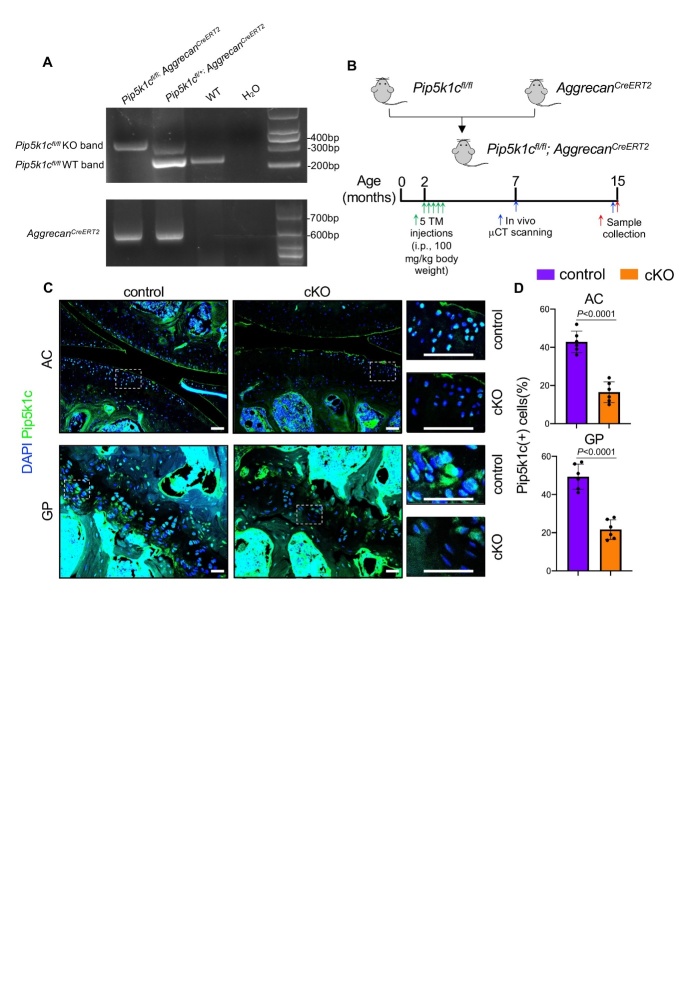
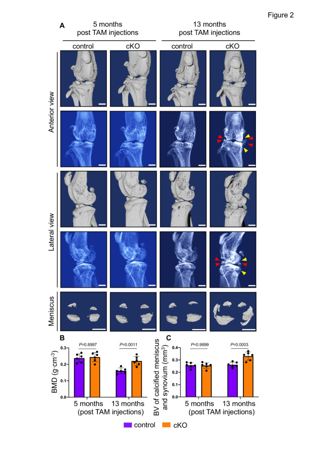
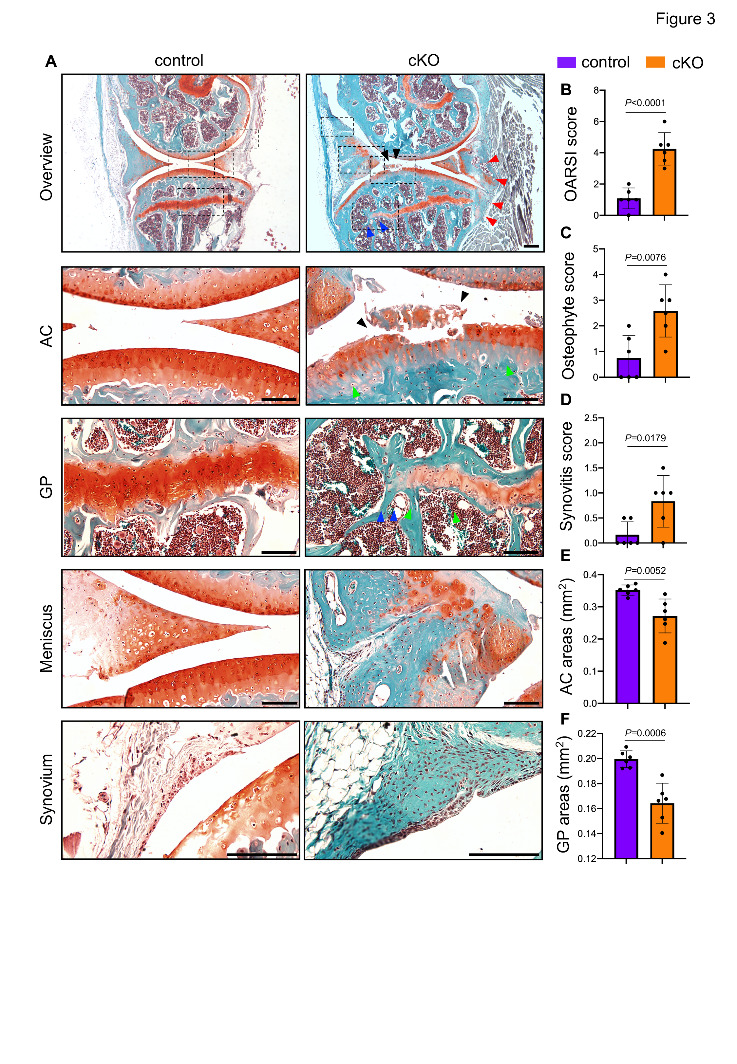
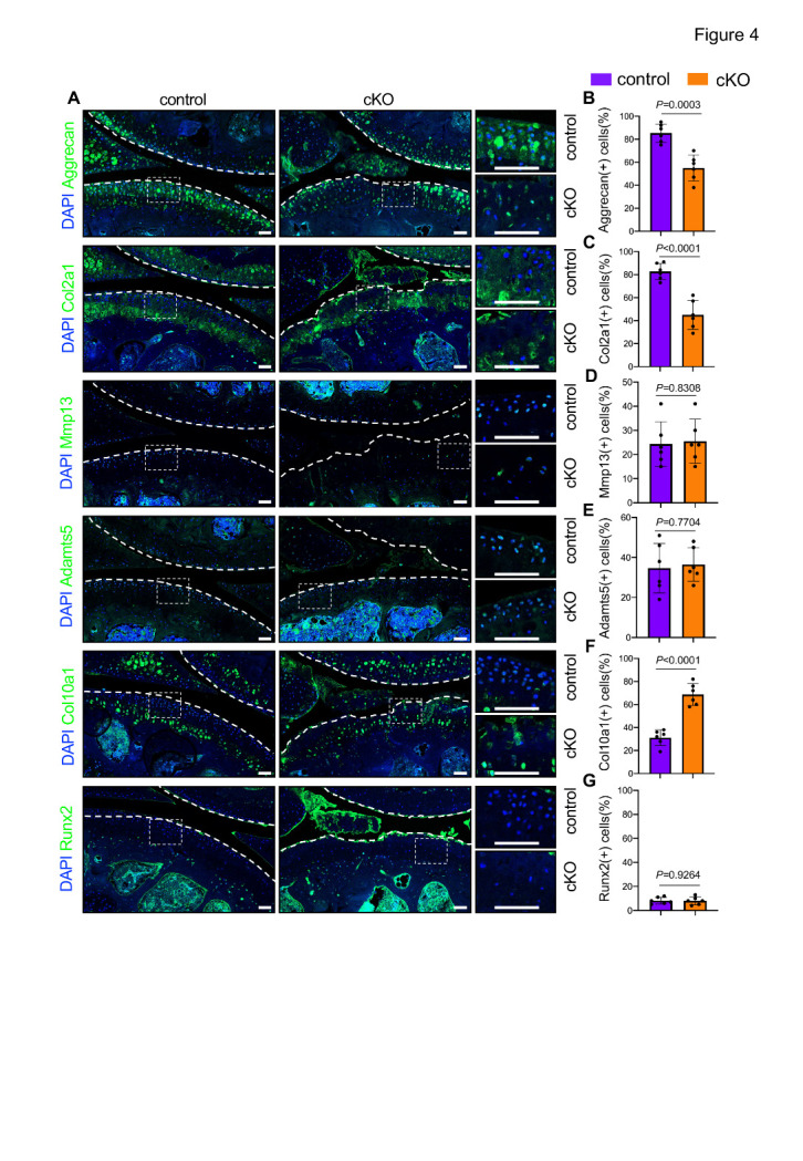
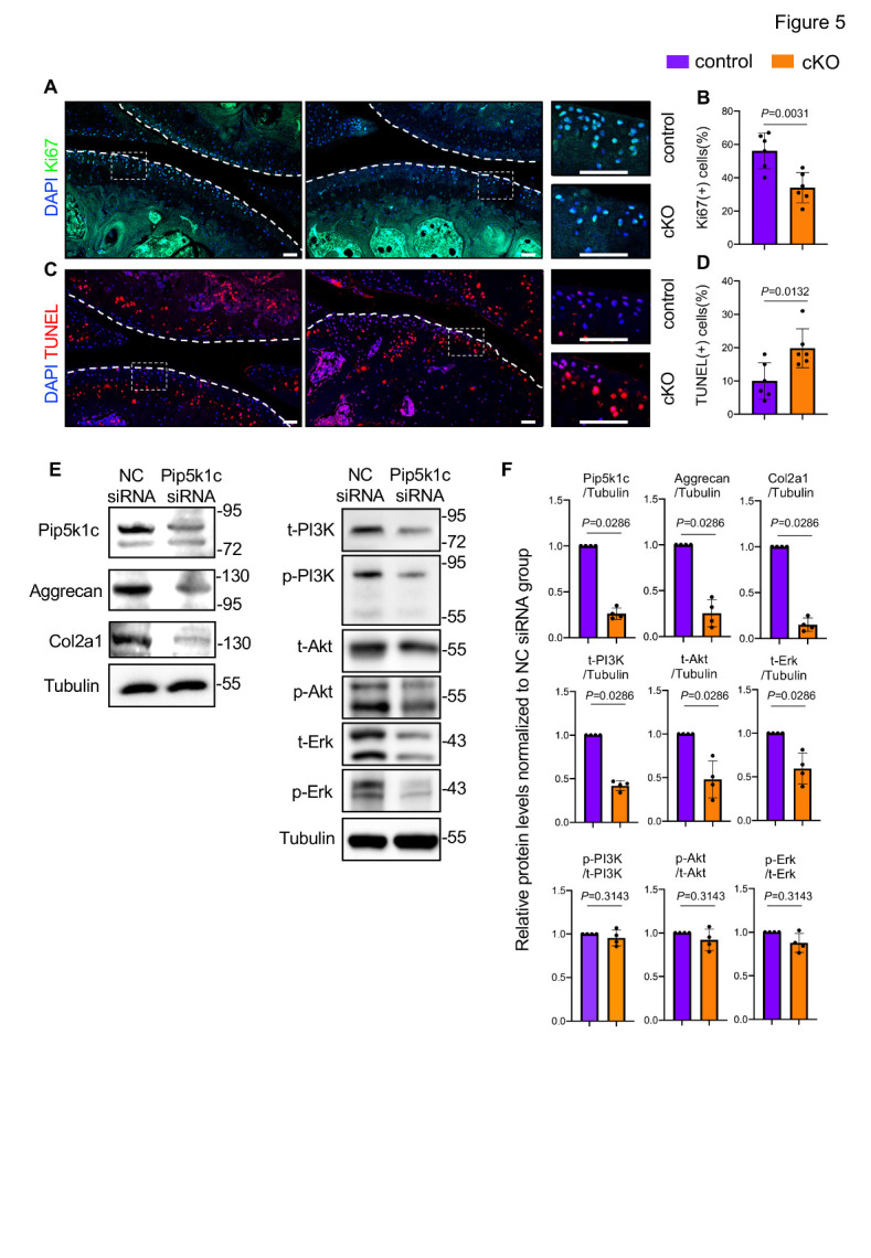
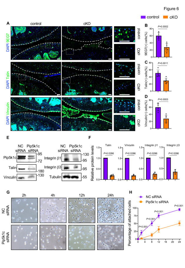
Osteoarthritis (OA) is the most common degenerative joint disease affecting the older populations globally. Phosphatidylinositol-4-phosphate 5-kinase type-1 gamma (Pip5k1c), a lipid kinase catalyzing the synthesis of phospholipid phosphatidylinositol 4,5-bisphosphate (PIP2), is involved in various cellular processes, such as focal adhesion (FA) formation, cell migration, and cellular signal transduction. However, whether Pip5k1c plays a role in the pathogenesis of OA remains unclear. Here we show that inducible deletion of Pip5k1c in aggrecan-expressing chondrocytes (cKO) causes multiple spontaneous OA-like lesions, including cartilage degradation, surface fissures, subchondral sclerosis, meniscus deformation, synovial hyperplasia, and osteophyte formation in aged (15-month-old) mice, but not in adult (7-month-old) mice. Pip5k1c loss promotes extracellular matrix (ECM) degradation, chondrocyte hypertrophy and apoptosis, and inhibits chondrocyte proliferation in the articular cartilage of aged mice. Pip5k1c loss dramatically downregulates the expressions of several key FA proteins, including activated integrin β1, talin, and vinculin, and thus impairs the chondrocyte adhesion and spreading on ECM. Collectively, these findings suggest that Pip5k1c expression in chondrocytes plays a critical role in maintaining articular cartilage homeostasis and protecting against age-related OA.
Keywords: Pip5k1c; aging; articular chondrocytes; osteoarthritis.
copyright: © 2022 Qu et al.
Conflict of interest statement
Conflicts of Interest The authors declare that they have no competing financial interests.
Figures
Similar articles
-
Kindlin-2 loss in condylar chondrocytes causes spontaneous osteoarthritic lesions in the temporomandibular joint in mice.Int J Oral Sci. 2022 Jul 4;14(1):33. doi: 10.1038/s41368-022-00185-1. Int J Oral Sci. 2022. PMID: 35788130 Free PMC article.
-
Molecular regulation of articular chondrocyte function and its significance in osteoarthritis.Histol Histopathol. 2011 Mar;26(3):377-94. doi: 10.14670/HH-26.377. Histol Histopathol. 2011. PMID: 21210351 Review.
-
Regulation of α5 and αV Integrin Expression by GDF-5 and BMP-7 in Chondrocyte Differentiation and Osteoarthritis.PLoS One. 2015 May 26;10(5):e0127166. doi: 10.1371/journal.pone.0127166. eCollection 2015. PLoS One. 2015. PMID: 26010756 Free PMC article.
-
Chondrocyte Apoptosis in the Pathogenesis of Osteoarthritis.Int J Mol Sci. 2015 Oct 30;16(11):26035-54. doi: 10.3390/ijms161125943. Int J Mol Sci. 2015. PMID: 26528972 Free PMC article. Review.
-
MiR-128-3p Post-Transcriptionally Inhibits WISP1 to Suppress Apoptosis and Inflammation in Human Articular Chondrocytes via the PI3K/AKT/NF-κB Signaling Pathway.Cell Transplant. 2020 Jan-Dec;29:963689720939131. doi: 10.1177/0963689720939131. Cell Transplant. 2020. PMID: 32830547 Free PMC article.
Cited by
-
Targeting Piezo1 channel to alleviate intervertebral disc degeneration.J Orthop Translat. 2025 Mar 8;51:145-158. doi: 10.1016/j.jot.2025.01.006. eCollection 2025 Mar. J Orthop Translat. 2025. PMID: 40129609 Free PMC article.
-
Pip5k1c expression in osteocytes regulates bone remodeling in mice.J Orthop Translat. 2024 Mar 8;45:36-47. doi: 10.1016/j.jot.2023.10.008. eCollection 2024 Mar. J Orthop Translat. 2024. PMID: 38495744 Free PMC article.
-
Integrin signalling in joint development, homeostasis and osteoarthritis.Nat Rev Rheumatol. 2024 Aug;20(8):492-509. doi: 10.1038/s41584-024-01130-8. Epub 2024 Jul 16. Nat Rev Rheumatol. 2024. PMID: 39014254 Free PMC article. Review.
-
Pip5k1γ promotes anabolism of nucleus pulposus cells and intervertebral disc homeostasis by activating CaMKII-Ampk pathway in aged mice.Aging Cell. 2024 Sep;23(9):e14237. doi: 10.1111/acel.14237. Epub 2024 Jun 5. Aging Cell. 2024. PMID: 38840443 Free PMC article.
-
Brief research report: Effects of Pinch deficiency on cartilage homeostasis in adult mice.Front Cell Dev Biol. 2023 Jan 19;11:1116128. doi: 10.3389/fcell.2023.1116128. eCollection 2023. Front Cell Dev Biol. 2023. PMID: 36743414 Free PMC article.
References
-
- Hunter DJ, Bierma-Zeinstra S (2019). Osteoarthritis. The Lancet, 393:1745-1759. - PubMed
-
- Glyn-Jones S, Palmer AJR, Agricola R, Price AJ, Vincent TL, Weinans H, et al.. (2015). Osteoarthritis. The Lancet, 386:376-387. - PubMed
-
- Safiri S, Kolahi AA, Smith E, Hill C, Bettampadi D, Mansournia MA, et al.. (2020). Global, regional and national burden of osteoarthritis 1990-2017: a systematic analysis of the Global Burden of Disease Study 2017. Ann Rheum Dis, 79:819-828. - PubMed
LinkOut - more resources
Full Text Sources