. 2023 Mar 16;8(12):11003-11020.
doi: 10.1021/acsomega.2c07762. eCollection 2023 Mar 28.

Quantitative Evaluation of a Multimodal Aptamer-Targeted Long-Circulating Polymer for Tumor Targeting

Affiliations

Quantitative Evaluation of a Multimodal Aptamer-Targeted Long-Circulating Polymer for Tumor Targeting

Lennart Bohrmann et al. ACS Omega. .

Abstract

Aptamers are promising targeting agents for imaging and therapy of numerous diseases, including cancer. However, a significant shortcoming of aptamers is their poor stability and fast excretion, limiting their application in vivo. Common strategies to overcome these challenges is to chemically modify aptamers in order to increase their stability and/or to apply formulation technologies such as conjugating them to polymers or nanocarriers in order to increase their circulation half-life. This is expected to result in improved cellular uptake or retention to passively targeted nanomedicines. Herein, we report a modular conjugation strategy based on click chemistry between functionalized tetrazines and trans-cyclooctene (TCO), for the modification of high molecular weight hyperbranched polyglycerol (HPG) with sgc8 aptamer, fluorescent dyes, and 111In. Our data indicate strong affinity of sgc8 against a range of solid tumor-derived cell lines that have previously not been tested with this aptamer. Nevertheless, nonspecific uptake of scrambled ssDNA-functionalized HPG in cells highlights inherent challenges of aptamer-targeted probes that remain to be solved for clinical translation. We validate HPG-sgc8 as a nontoxic nanoprobe with high affinity against MDA-MB-468 breast and A431 lung cancer cells and show significantly increased plasma stability compared to free sgc8. In vivo quantitative SPECT/CT imaging indicates EPR-mediated tumor uptake of HPG-sgc8 and nontargeted or scrambled ssDNA-conjugated HPG but no statistically significant difference between these formulations in terms of total tumor uptake or retention. Our study emphasizes the need for stringent controls and quantification in the evaluation of aptamer-targeted probes. For this purpose, our versatile synthesis strategy provides a simple approach for the design and evaluation of long-circulating aptamer-conjugated nanoformulations.


Conflict of interest statement

The authors declare no competing financial interest.

Figures

Scheme 1
Scheme 1. Functionalization of HPG with TCO and Subsequent Conjugation of ssDNA (sgc8/scr), Fluorescent Dyes (Cy3/Cy5), and the Radioisotope 111In
The primary amines on HPG are TCO-functionalized via activated ester conjugation with TCO-PEG4-NHS. A variety of ssDNA, fluorescence, and 111In-DTPA-modified tetrazines is clicked to HPG-TCO using IEDDA click chemistry to afford a targeted multimodal fluorescence and SPECT nanoprobe.
Figure 1
Figure 1
In vitro characterization of HPG-ssDNA constructs. (A) TBE-Urea gel electrophoresis of [111In]In-HPG-sgc8 (1), [111In]In-HPG-scr (2), NH2-sgc8 (3), and NH2-scr (4). SYBR Gold was used to stain ssDNA. The radioactive signal of 111In was recorded on a phosphor imager. (B) TBE-Urea gel electrophoresis of HPG-sgc8-Cy5 (1) and HPG-scr-Cy5 (2). SYBR Gold was used to stain ssDNA; the red signal in the Cy5 channel corresponds to the fluorophore bound to HPG. (C) Radio-HPLC chromatograms of fresh [111In]InCl3 (blue), the labeled tetrazine prosthetic group [111In]In-Tz-DTPA used for functionalization (red), and purified [111In]In-HPG-sgc8 (black). (D) Hydrodynamic size and ζ potential of HPG-NH2, HPG-TCO, HPG-Cy3, HPG-sgc8-Cy3, and HPG-scr-Cy3. Note that Cy3 was used to avoid interference with the laser of the DLS measurement. Conjugation of ssDNA leads to slightly increased size of the construct and a more negative ζ potential, corresponding to the negatively charged ssDNA.
Figure 2
Figure 2
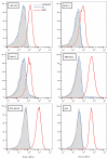
Binding of sgc8 aptamer to various cell lines. AlexaFluor647-labeled sgc8 was incubated with cells at a concentration of 5 nM. Strong binding was observed for MIA PaCa, MDA-MB-468, and A431 cells, while moderate binding was observed for HEK-293, BxPC-3, and Capan-2 cells. No binding was observed for a scrambled control sequence (AlexaFluor647-scr).
Figure 3
Figure 3
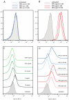
ssDNA-mediated specific binding of HPG against HEK-293 (A, B) and MDA-MB-468 cells (C, D). (A) No binding of HPG-scr at concentrations of 0.1 to 10 nM is observed. (B) Strong binding is observed for HPG-sgc8 even at low-nanomolar concentrations. (C) Nonfunctionalized HPG does not show nonspecific binding until high concentrations. (D) ssDNA-modified HPG shows specific binding for sgc8 even at low to moderate concentrations. Nonspecific binding of HPG-scr is observed only at a concentration of 500 nM.
Figure 4
Figure 4
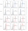
Effect of temperature on specific and nonspecific binding of HPG-ssDNA on A431 cells. At 4 °C, binding saturation of HPG-Cy5-sgc8 was reached at a concentration of 1 nM and did not increase further with higher concentrations. No binding was observed for HPG-Cy5-scr over the same concentration range. A similar profile for HPG-sgc8 was observed at 37 °C, while HPG-Cy5-scr showed considerable concentration-dependent uptake. Nontreated cells are shown in light gray; colored lines are cells treated with HPG-Cy5-sgc8 and HPG-Cy5-scr, respectively.
Figure 5
Figure 5
Confocal imaging of HPG-Cy5-sgc8 and HPG-Cy5-scr on HEK-293, MDA-MB-468 and A431 cells at 40× magnification. HPG-Cy5-sgc8 shows clear membrane localization with some diffuse intracellular uptake, especially in MDA-MB-468 cells. HPG-Cy5-scr does not bind to the cell membrane, and only weak intracellular uptake is visible. The scale bar in all pictures is 50 μm.
Figure 6
Figure 6
Cell binding of [111In]In-HPG-sgc8 and in vitro pretargeted labeling of HPG-sgc8 using tetrazine-Cy3 and the [111In]In prosthetic group. (A) MDA-MB-468 cells incubated with HPG-Cy5-sgc8. After 30 min tetrazine Cy3 was added before the cells were stained with fixing medium containing DAPI. Obvious membrane staining indicates binding of the nanoprobe to PTK7 on the cell surface; a more diffuse uptake points to some internalization. Tetrazine-Cy3 is only able to bind to free TCO groups on the membrane-bound HPG. The scale bar in the pictures is 25 μm. (B) Binding affinity of 111In-labeled HPG-sgc8 to MDA-MB-468 cells. (C) Cells preincubated with HPG-sgc8 showing a more than 2-fold increased binding of [111In]In-Tz-DTPA compared to cells treated with HPG-scr or PBS. Statistical significance (*, p < 0.05) was determined using Student’s t test; n = 3.
Figure 7
Figure 7
Cellular toxicity of aptamers and HPG functionalized with aptamers. (A) Proliferation of HUVEC cells in medium supplemented with free sgc8 or scr aptamer. No effect on growth of HUVEC (B), MDA-MB-468 (C), and HEK-293 (D) cells incubated with HPG-sgc8 and HPG-scr was observed.
Figure 8
Figure 8
Stability of sgc8 and HPG-sgc8 in plasma. Sgc8 and HPG-sgc8 was incubated in fresh mouse plasma at 37 °C. At different time points aliquots were taken and analyzed using TBE-urea gel electrophoresis. DNA was visualized using SYBR Gold nucleic acid stain. The intensity was normalized to time 0 and fitted using a one-phase exponential decay function.
Figure 9
Figure 9
In vivo imaging of HPG-aptamer probes. (A) SPECT/CT MIP rendering of [111In]In-HPG-Cy5-sgc8. Representative rendering of a mouse bearing a subcutaneous A431 tumor on the lower back. Initially high blood levels of the nanoprobe are visible, indicated by activity in carotid arteries, heart, and lungs. Liver uptake is evident at all time points but increases over time. Bladder activity is only visible at the earliest time points, although kidney levels remain relatively high throughout the study. Spleen uptake is visible after 48 h. CT in gray scale, SPECT in color. B, bladder; C, carotid artery; H, heart; K, kidney; L, lung; Li, liver; S, spleen; T, tumor area indicated by dashed circle. (B–D) Mean SUV in g/mL for blood pool, liver, kidney, and bladder. The majority of the activity for all formulations remained in the body; small amounts were excreted via renal and hepatobiliary elimination. (E) Comparison of SUVmax and SUV95th in tumor region. [111In]In-HPG-Cy5-scr shows consistently lower SUVmax in the tumor region, directly after injection, [111In]In-HPG-Cy5-sgc8 shows a statistically significant higher SUVmax than the other formulations. (F) %ID in tumor region over time. [111In]In-HPG-Cy5-scr shows a lower uptake than [111In]In-HPG-Cy5-sgc8 and [111In]In-HPG-Cy5. (G) Postmortem biodistribution (%ID/organ) at 48 h postinjection. Notable uptake was observed in blood, liver, kidneys, and tumors. Data represented as mean ± SD; n = 3. Statistical significance (*, p < 0.05) was determined using two-way repeated measurement ANOVA with a Holm–Sidak correction.
Figure 10
Figure 10
Representative tumor histology of HPG-Cy5-sgc8 injected mice. (A) Gross overview of A431 tumor. Large cystic core showing necrotic cells and void areas that were lost during sample preparation. Close ups of (B) tumor periphery, (C) necrotic area, and (D) central viable tumor region. Confocal images showing cell nuclei counterstained with DAPI in blue and the location of HPG-Cy5-sgc8 in red. (E) Majority of formulation found in the periphery of the tumor. (F) Very little fluorescence found in the core of the tumor. Spleen (G) and liver (H) showing high uptake of HPG-Cy5-sgc8, pointing to uptake by the reticuloendothelial system.

Similar articles

Cited by

References

    1. Lee D.-E.; Koo H.; Sun I.-C.; Ryu J. H.; Kim K.; Kwon I. C. Multifunctional nanoparticles for multimodal imaging and theragnosis. Chem. Soc. Rev. 2012, 41 (7), 2656–2672. 10.1039/C2CS15261D. - DOI - PubMed
    1. Key J.; Leary J. F. Nanoparticles for multimodal in vivo imaging in nanomedicine. Int. J. Nanomed. 2014, 9, 711–726. 10.2147/IJN.S53717. - DOI - PMC - PubMed
    1. Chapman S.; Dobrovolskaia M.; Farahani K.; Goodwin A.; Joshi A.; Lee H.; Meade T.; Pomper M.; Ptak K.; Rao J.; Singh R.; Sridhar S.; Stern S.; Wang A.; Weaver J. B.; Woloschak G.; Yang L. Nanoparticles for cancer imaging: The good, the bad, and the promise. Nano Today 2013, 8 (5), 454–460. 10.1016/j.nantod.2013.06.001. - DOI - PMC - PubMed
    1. Reisch A.; Klymchenko A. S. Fluorescent Polymer Nanoparticles Based on Dyes: Seeking Brighter Tools for Bioimaging. Small 2016, 12 (15), 1968–1992. 10.1002/smll.201503396. - DOI - PMC - PubMed
    1. Kainthan R. K.; Muliawan E. B.; Hatzikiriakos S. G.; Brooks D. E. Synthesis, Characterization, and Viscoelastic Properties of High Molecular Weight Hyperbranched Polyglycerols. Macromolecules 2006, 39 (22), 7708–7717. 10.1021/ma0613483. - DOI