Exosomes from adipose-derived stem cells promote angiogenesis and reduce necrotic grade in hindlimb ischemia mouse models
- PMID: 37009008
- PMCID: PMC10008393
- DOI: 10.22038/IJBMS.2023.67936.14857
Exosomes from adipose-derived stem cells promote angiogenesis and reduce necrotic grade in hindlimb ischemia mouse models
Abstract
Objectives: Acute hindlimb ischemia is a peripheral arterial disease that severely affects the patient's health. Injection of stem cells-derived exosomes that promote angiogenesis is a promising therapeutic strategy to increase perfusion and repair ischemic tissues. This study aimed to evaluate the efficacy of adipose stem cell-derived exosomes injection (ADSC-Exos) in treating acute mouse hindlimb ischemia.
Materials and methods: ADSC-Exos were collected via ultracentrifugation. Exosome-specific markers were analyzed via flow cytometry. The morphology of exosomes was detected by TEM. A dose of 100 ug exosomes/100 ul PBS was locally injected into acute mice ischemic hindlimb. The treatment efficacy was evaluated based on the oxygen saturation level, limb function, new blood vessel formation, muscle structure recovery, and limb necrosis grade.
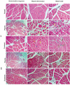
Results: ADSC-exosomes expressed high positivity for markers CD9 (76.0%), CD63 (91.2%), and CD81 (99.6%), and have a cup shape. After being injected into the muscle, in the treatment group, many small and short blood vessels formed around the first ligation and grew down toward the second ligation. The SpO2 level, reperfusion, and recovery of the limb function are more positively improved in the treatment group. On day 28, the muscle's histological structure in the treatment group is similar to normal tissue. Approximately 33.33% of the mice had grade I and II lesions and there were no grade III and IV observed in the treatment group. Meanwhile, in the placebo group, 60% had grade I to IV lesions.
Conclusion: ADSC-Exos showed the ability to stimulate angiogenesis and significantly reduce the rate of limb necrosis.
Keywords: Acute; Adipose-derived stem cells; Angiogenesis; Exosome; Extracellular vesicles; Limb ischemia.
Conflict of interest statement
The authors declare that they have no competing interests.
Figures


Similar articles
-
Intravenous Infusion of Exosomes Derived from Human Adipose Tissue-Derived Stem Cells Promotes Angiogenesis and Muscle Regeneration: An Observational Study in a Murine Acute Limb Ischemia Model.Adv Exp Med Biol. 2023 Mar 30. doi: 10.1007/5584_2023_769. Online ahead of print. Adv Exp Med Biol. 2023. PMID: 36991295
-
Exosomal miR-125b-5p derived from adipose-derived mesenchymal stem cells enhance diabetic hindlimb ischemia repair via targeting alkaline ceramidase 2.J Nanobiotechnology. 2023 Jun 12;21(1):189. doi: 10.1186/s12951-023-01954-8. J Nanobiotechnology. 2023. PMID: 37308908 Free PMC article.
-
Adipose-derived stem cell-secreted exosomes enhance angiogenesis by promoting macrophage M2 polarization in type 2 diabetic mice with limb ischemia via the JAK/STAT6 pathway.Heliyon. 2022 Nov 13;8(11):e11495. doi: 10.1016/j.heliyon.2022.e11495. eCollection 2022 Nov. Heliyon. 2022. PMID: 36406687 Free PMC article.
-
Adipose-Derived Stem Cell Exosomes as a Novel Anti-Inflammatory Agent and the Current Therapeutic Targets for Rheumatoid Arthritis.Biomedicines. 2022 Jul 18;10(7):1725. doi: 10.3390/biomedicines10071725. Biomedicines. 2022. PMID: 35885030 Free PMC article. Review.
-
Therapeutic potential of exosomes from adipose-derived stem cells in chronic wound healing.Front Surg. 2022 Sep 30;9:1030288. doi: 10.3389/fsurg.2022.1030288. eCollection 2022. Front Surg. 2022. PMID: 36248361 Free PMC article. Review.
Cited by
-
The Dual Effects of Exosomes on Glioma: A Comprehensive Review.J Cancer. 2023 Sep 4;14(14):2707-2719. doi: 10.7150/jca.86996. eCollection 2023. J Cancer. 2023. PMID: 37779868 Free PMC article. Review.
-
Intramyocardial injection of pre-cultured endothelial progenitor cells and mesenchymal stem cells inside alginate/gelatin microspheres induced angiogenesis in infarcted rabbits.Cell Commun Signal. 2025 Jun 12;23(1):279. doi: 10.1186/s12964-025-02301-0. Cell Commun Signal. 2025. PMID: 40506721 Free PMC article.
-
Emerging Strategies for Revascularization: Use of Cell-Derived Extracellular Vesicles and Artificial Nanovesicles in Critical Limb Ischemia.Bioengineering (Basel). 2025 Jan 20;12(1):92. doi: 10.3390/bioengineering12010092. Bioengineering (Basel). 2025. PMID: 39851366 Free PMC article. Review.
-
Angiogenic Ability of Extracellular Vesicles Derived from Angio-miRNA-Modified Mesenchymal Stromal Cells.Tissue Eng Regen Med. 2025 Jul 31. doi: 10.1007/s13770-025-00741-w. Online ahead of print. Tissue Eng Regen Med. 2025. PMID: 40742422
-
Biomaterial-based vascularization strategies for enhanced treatment of peripheral arterial disease.J Nanobiotechnology. 2025 Feb 12;23(1):103. doi: 10.1186/s12951-025-03140-4. J Nanobiotechnology. 2025. PMID: 39940018 Free PMC article. Review.
References
-
- Martyn Knowles CHT. Epidemiology of acute critical limb ischemia. Critical Limb Ischemia. Cham: Springer; 2017. pp. 1–7.
-
- Lawall H, Huppert P, Rümenapf G. S3-Leitlinie zur diagnostik, therapie und nachsorge der peripheren arteriellen verschlusskrankheit. Vasa. 2016;45:11–82. - PubMed
-
- Fluck F, Augustin AM, Bley T, Kickuth R. Current treatment options in acute limb ischemia. Rofo. 2020;192:319–326. - PubMed
LinkOut - more resources
Full Text Sources
Miscellaneous
