. 2023 Feb 13;4(3):287-298.
doi: 10.1021/accountsmr.2c00239. eCollection 2023 Mar 24.

Complex Coacervate Materials as Artificial Cells

Affiliations

Complex Coacervate Materials as Artificial Cells

Alexander B Cook et al. Acc Mater Res. .

Abstract

Cells have evolved to be self-sustaining compartmentalized systems that consist of many thousands of biomolecules and metabolites interacting in complex cycles and reaction networks. Numerous subtle intricacies of these self-assembled structures are still largely unknown. The importance of liquid-liquid phase separation (both membraneless and membrane bound) is, however, recognized as playing an important role in achieving biological function that is controlled in time and space. Reconstituting biochemical reactions in vitro has been a success of the last decades, for example, establishment of the minimal set of enzymes and nutrients able to replicate cellular activities like the in vitro transcription translation of genes to proteins. Further than this though, artificial cell research has the aim of combining synthetic materials and nonliving macromolecules into ordered assemblies with the ability to carry out more complex and ambitious cell-like functions. These activities can provide insights into fundamental cell processes in simplified and idealized systems but could also have an applied impact in synthetic biology and biotechnology in the future. To date, strategies for the bottom-up fabrication of micrometer scale life-like artificial cells have included stabilized water-in-oil droplets, giant unilamellar vesicles (GUV's), hydrogels, and complex coacervates. Water-in-oil droplets are a valuable and easy to produce model system for studying cell-like processes; however, the lack of a crowded interior can limit these artificial cells in mimicking life more closely. Similarly membrane stabilized vesicles, such as GUV's, have the additional membrane feature of cells but still lack a macromolecularly crowded cytoplasm. Hydrogel-based artificial cells have a macromolecularly dense interior (although cross-linked) that better mimics cells, in addition to mechanical properties more similar to the viscoelasticity seen in cells but could be seen as being not dynamic in nature and limiting to the diffusion of biomolecules. On the other hand, liquid-liquid phase separated complex coacervates are an ideal platform for artificial cells as they can most accurately mimic the crowded, viscous, highly charged nature of the eukaryotic cytoplasm. Other important key features that researchers in the field target include stabilizing semipermeable membranes, compartmentalization, information transfer/communication, motility, and metabolism/growth. In this Account, we will briefly cover aspects of coacervation theory and then outline key cases of synthetic coacervate materials used as artificial cells (ranging from polypeptides, modified polysaccharides, polyacrylates, and polymethacrylates, and allyl polymers), finishing with envisioned opportunities and potential applications for coacervate artificial cells moving forward.


Conflict of interest statement

The authors declare no competing financial interest.

Figures
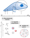
Figure 1
Figure 1
General schematic showing approaches to fabricate coacervate-based artificial cell systems, i) (macro)molecular materials used for both liquid–liquid phase separated coacervate droplets and membranes, ii) strategies to include subcellular compartments through further phase separation or inclusion of polymersomes or other nanoscale particles, and iii) assembly of all components to form micrometer-sized hierarchical coacervate-based artificial cells.
Figure 2
Figure 2
Schematic overview representing liquid–liquid phase separation of macromolecular solutions. a) Representation of associative and segregative coacervation, on the macromolecular level, with corresponding simplified phase diagrams showing one phase regions, two phase regions and tie-lines. b) Selected chemical structures of synthetic macromolecules applied as complex coacervate-based artificial cell systems.
Figure 3
Figure 3
Synthetic block copolymer membrane PEG-(PCLgTMC)-PGlu and its assembly on the surface of liquid–liquid phase separated droplets, as artificial cell models. A) Confocal micrograph showing coacervate core and membrane interface. B) 3D model representation of assembled structure. Reproduced with permission from ref (19). Copyright 2017 American Chemical Society. Distributed under a Creative Commons Attribution Non-Commercial No Derivative Works 4.0 Usage Agreement.
Figure 4
Figure 4
Polypeptide-based multiphase coacervates with phase specific RNA accumulation. Chemical structures of peptides studied and Förster Resonance Energy Transfer (FRET) microscopy images with RNA duplex 10mer with each strand bearing either Cy3 or Cy5 fluorophore. a) Overall thermodynamic equilibria schematic showing the proposed dynamics of the system. b), c) Quantification of partitioning thermodynamic parameters from FRET data analysis. d), e) Quantification of dissociation thermodynamic parameters from FRET data analysis. Reproduced with permission from ref (15). Copyright 2022 The Authors.
Figure 5
Figure 5
a) Synthetic polymers for the recruitment of small molecules and biomolecules. b) Partitioning coefficients of rhodamine B and BSA. c) Apparent polarity of coacervate droplets with respect to water. d) Fusion times of various polymer droplets. e), f) Cloud point of coacervate droplets varied by monomer composition. g) Salt dependence of FITC-BSA uptake and coacervate disassembly. Reproduced with permission from ref (26). Copyright 2021 The Author(s) distributed under a Creative Commons Attribution License 4.0 (CC BY-NC).
Figure 6
Figure 6
Protein-based shuttling between different populations of complex coacervate artificial cells. First population containing eYFP and a specific DNA strand, upon addition (and coacervate uptake) of a complementary DNA strand, the protein is excreted. eYFP is then taken up by the second population of artificial cells, as observed by confocal microscopy over 60 min. Reproduced with permission from ref (47). Copyright 2022 The Author(s).
Figure 7
Figure 7
Enzyme decorated coacervate artificial cells able to process multiple signaling molecules. a) Formation of lipid membrane stabilized PDDA/DNA coacervate droplets and enzyme immobilization to achieve. b) Gox, HRP, and CAT coacervate droplets. c) Bright-field. d) Fluorescence. e) Optical phase contrast. f) Dark field microscopy images of complex coacervates showing continuous phospholipid membrane. g) Fabrication of tubular prototissue containing three artificial cell populations embedded in hydrogel. h), (i) Enzyme cascade reaction schematic showing positions of H2O2 and NO monitoring (i–iv) and substrate reaction diffusion process, j), k) H2O2 and NO were monitored over time with colorimetric reactions (ABTS oxidation, and Greiss reagent). Reproduced with permission from ref (50). Copyright 2022 The Author(s) distributed under a Creative Commons Attribution License 4.0 (CC BY-NC).
Figure 8
Figure 8
Enzyme motor driven motility of polymer coacervate droplets. a) Schematic diagram of transient asymmetry in enzyme distribution due to lateral diffusion in the membrane. b) Trajectories of urease or c) catalase motile artificial cells, fueled by the addition of urea or H2O2, respectively. d) Mean square displacement curves of urease and e) catalase motile artificial cells. Reproduced with permission from ref (53). Copyright 2021 The Author(s) distributed under a Creative Commons Attribution-Noncommercial License 4.0 (CC BY-NC).
Figure 9
Figure 9
Assembly of live-cell containing polymer coacervate system and its dynamic morphogenesis. a) Confocal microscopy images over time showing transformation from spherical artificial cells to nonspherical. b) 3D reconstruction. c) Changes in number of live E. coli cells per artificial cell. d) Mean relative volumes of artificial cells over time. e) Diagram. f) Confocal micrograph of nonspherical artificial cell showing life-like features: (1) outer membrane, (2) crowded macromolecular cytoplasm, (3) DNA/Histone organelle, (4) encapsulated E. coli (mitochondria mimicking), (5) actin network, (6) spherical vacuoles, (7) amoeba-like morphology. Scale bars: 10 μm. Reproduced with permission from ref (14). Copyright 2022 The Authors.

References

    1. Buddingh’ B. C.; van Hest J. C. M. Artificial Cells: Synthetic Compartments with Life-like Functionality and Adaptivity. Acc. Chem. Res. 2017, 50 (4), 769–777. 10.1021/acs.accounts.6b00512. - DOI - PMC - PubMed
    1. Qian X.; Nymann Westensee I.; Brodszkij E.; Städler B. Cell Mimicry as a Bottom-up Strategy for Hierarchical Engineering of Nature-Inspired Entities. WIREs Nanomedicine Nanobiotechnology 2021, 13 (3), e168310.1002/wnan.1683. - DOI - PubMed
    1. van Stevendaal M. H. M. E.; van Hest J. C. M.; Mason A. F. Functional Interactions Between Bottom-Up Synthetic Cells and Living Matter for Biomedical Applications. ChemSystemsChem. 2021, 3 (5), e2100009.10.1002/syst.202100009. - DOI
    1. Hyman A. A.; Weber C. A.; Jülicher F. Liquid-Liquid Phase Separation in Biology. Annu. Rev. Cell Dev. Biol. 2014, 30 (1), 39–58. 10.1146/annurev-cellbio-100913-013325. - DOI - PubMed
    1. Insua I.; Montenegro J. Synthetic Supramolecular Systems in Life-like Materials and Protocell Models. Chem. 2020, 6 (7), 1652–1682. 10.1016/j.chempr.2020.06.005. - DOI