

Early treatment with dapsone after spinal cord injury in rats decreases the inflammatory response and promotes long-term functional recovery
- PMID: 37009237
- PMCID: PMC10060111
- DOI: 10.1016/j.heliyon.2023.e14687
Early treatment with dapsone after spinal cord injury in rats decreases the inflammatory response and promotes long-term functional recovery
Abstract
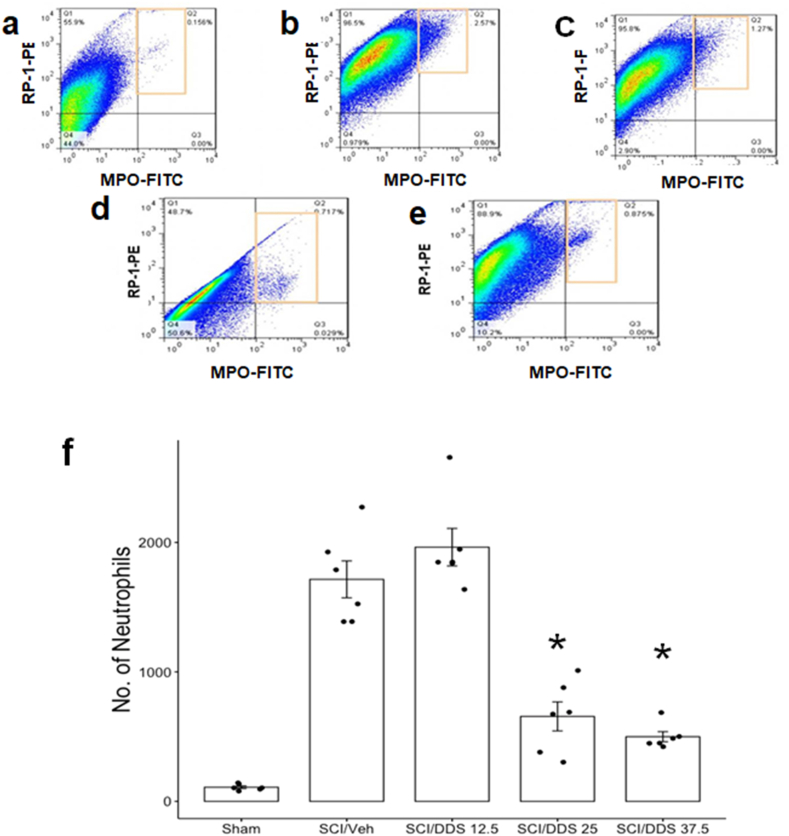
Failure of therapeutic strategies for the management and recovery from traumatic spinal cord injury (SCI) is a serious concern. Dapsone (DDS) has been reported as a neuroprotective drug after SCI, although the phase after SC damage (acute or chronic) of its major impact on functional recovery has yet to be defined. Here, we evaluated DDS acute-phase anti-inflammatory effects and their impact on early functional recovery, one week after moderate SCI, and late functional recovery, 7 weeks thereafter. Female Wistar rats were randomly assigned to each of five experimental groups: sham group; four groups of rats with SCI, treated with DDS (0, 12.5, 25.0, and 37.5 mg/kg ip), starting 3 h after injury. Plasma levels of GRO/KC, and the number of neutrophils and macrophages in cell suspensions from tissue taken at the site of injury were measured as inflammation biomarkers. Hindlimb motor function of injured rats given DDS 12.5 and 25.0 mg/kg daily for 8 weeks was evaluated on the BBB open-field ordinal scale. Six hours after injury all DDS doses decreased GRO/KC plasma levels; 24 h after injury, neutrophil numbers decreased with DDS doses of 25.0 and 37.5 mg/kg; macrophage numbers decreased only at the 37.5 mg/kg dose. In the acute phase, functional recovery was dose-dependent. Final recovery scores were 57.5 and 106.2% above the DDS-vehicle treated control group, respectively. In conclusion, the acute phase dose-dependent anti-inflammatory effects of DDS impacted early motor function recovery affecting final recovery at the end of the study.
Keywords: Dapsone; Inflammatory response; Interleukin-8; Motor function recovery; Spinal cord injury.
© 2023 The Authors.
Conflict of interest statement
The authors declare that they have no known competing financial interests or personal relationships that could have appeared to influence the work reported in this paper.
Figures


Similar articles
-
Timing and duration of anti-alpha4beta1 integrin treatment after spinal cord injury: effect on therapeutic efficacy.J Neurosurg Spine. 2009 Nov;11(5):575-87. doi: 10.3171/2009.6.SPINE08915. J Neurosurg Spine. 2009. PMID: 19929361
-
Delayed administration of dapsone protects from tissue damage and improves recovery after spinal cord injury.J Neurosci Res. 2011 Mar;89(3):373-80. doi: 10.1002/jnr.22555. Epub 2011 Jan 6. J Neurosci Res. 2011. PMID: 21259324
-
Delayed administration of high dose human immunoglobulin G enhances recovery after traumatic cervical spinal cord injury by modulation of neuroinflammation and protection of the blood spinal cord barrier.Neurobiol Dis. 2021 Jan;148:105187. doi: 10.1016/j.nbd.2020.105187. Epub 2020 Nov 26. Neurobiol Dis. 2021. PMID: 33249350
-
[Effects of curcumin on the recovery of hind limb function after spinal cord injury in rats and its mechamism].Zhongguo Ying Yong Sheng Li Xue Za Zhi. 2017 May 8;33(5):441-444. doi: 10.12047/j.cjap.5548.2017.106. Zhongguo Ying Yong Sheng Li Xue Za Zhi. 2017. PMID: 29926590 Chinese.
-
Anti-Apoptotic Effects of Dapsone After Spinal Cord Injury in Rats.Neurochem Res. 2015 Jun;40(6):1243-51. doi: 10.1007/s11064-015-1588-z. Epub 2015 May 1. Neurochem Res. 2015. PMID: 25931161
Cited by
-
Acute anticonvulsant effects of dapsone on PTZ- and MES-induced seizures in mice: NLRP3 inflammasome inhibition and Nrf2/HO-1 pathway preservation.Pharmacol Rep. 2025 Apr;77(2):450-462. doi: 10.1007/s43440-025-00698-6. Epub 2025 Jan 27. Pharmacol Rep. 2025. PMID: 39869286
-
The OSR9 Regimen: A New Augmentation Strategy for Osteosarcoma Treatment Using Nine Older Drugs from General Medicine to Inhibit Growth Drive.Int J Mol Sci. 2023 Oct 23;24(20):15474. doi: 10.3390/ijms242015474. Int J Mol Sci. 2023. PMID: 37895152 Free PMC article. Review.
-
IPIAD- an augmentation regimen added to standard treatment of pancreatic ductal adenocarcinoma using already-marketed repurposed drugs irbesartan, pyrimethamine, itraconazole, azithromycin, and dapsone.Oncoscience. 2024 Feb 7;11:15-31. doi: 10.18632/oncoscience.594. eCollection 2024. Oncoscience. 2024. PMID: 38524376 Free PMC article.
-
Investigating the efficacy of dapsone in treating sepsis induced by cecal ligation and puncture surgery in male mice.Naunyn Schmiedebergs Arch Pharmacol. 2024 Dec;397(12):9909-9917. doi: 10.1007/s00210-024-03251-z. Epub 2024 Jun 28. Naunyn Schmiedebergs Arch Pharmacol. 2024. PMID: 38940849
References
-
- Oyinbo C.A. Secondary injury mechanisms in traumatic spinal cord injury: a nugget of this multiply cascade. Acta Neurobiol. Exp. (Wars) 2011;71:281–299. - PubMed
LinkOut - more resources
Full Text Sources
