. 2023 Apr;55(4):767-778.
doi: 10.1038/s12276-023-00958-6. Epub 2023 Apr 3.

Evogliptin, a DPP-4 inhibitor, prevents diabetic cardiomyopathy by alleviating cardiac lipotoxicity in db/db mice

Affiliations

Evogliptin, a DPP-4 inhibitor, prevents diabetic cardiomyopathy by alleviating cardiac lipotoxicity in db/db mice

Trong Kha Pham et al. Exp Mol Med. 2023 Apr.

Abstract

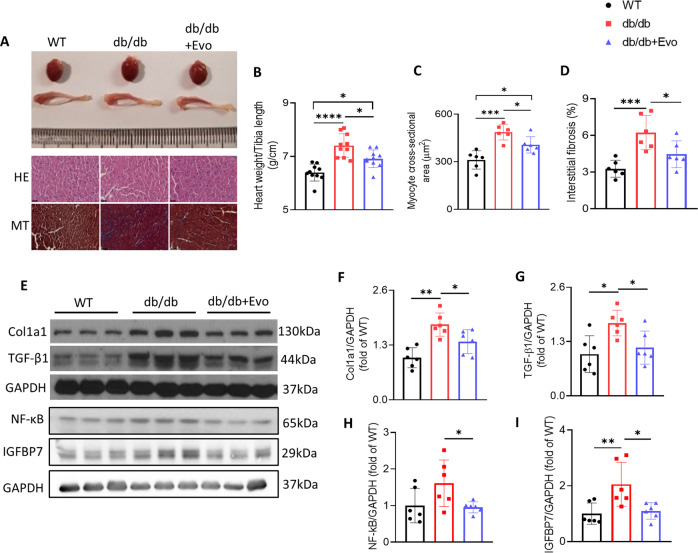
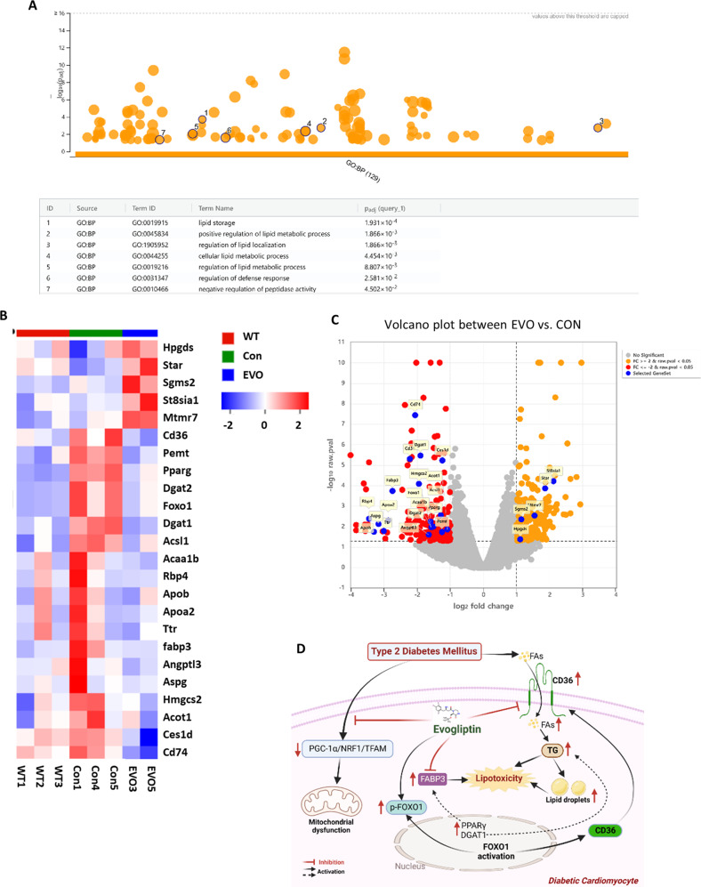
Dipeptidyl peptidase-4 (DPP-4) inhibitors are glucose-lowering drugs for type 2 diabetes mellitus (T2DM). We investigated whether evogliptin® (EVO), a DPP-4 inhibitor, could protect against diabetic cardiomyopathy (DCM) and the underlying mechanisms. Eight-week-old diabetic and obese db/db mice were administered EVO (100 mg/kg/day) daily by oral gavage for 12 weeks. db/db control mice and C57BLKS/J as wild-type (WT) mice received equal amounts of the vehicle. In addition to the hypoglycemic effect, we examined the improvement in cardiac contraction/relaxation ability, cardiac fibrosis, and myocardial hypertrophy by EVO treatment. To identify the mechanisms underlying the improvement in diabetic cardiomyopathy by EVO treatment, its effect on lipotoxicity and the mitochondrial damage caused by lipid droplet accumulation in the myocardium were analyzed. EVO lowered the blood glucose and HbA1c levels and improved insulin sensitivity but did not affect the body weight or blood lipid profile. Cardiac systolic/diastolic function, hypertrophy, and fibrosis were improved in the EVO-treated group. EVO prevented cardiac lipotoxicity by reducing the accumulation of lipid droplets in the myocardium through suppression of CD36, ACSL1, FABP3, PPARgamma, and DGAT1 and enhancement of the phosphorylation of FOXO1, indicating its inhibition. The EVO-mediated improvement in mitochondrial function and reduction in damage were achieved through activation of PGC1a/NRF1/TFAM, which activates mitochondrial biogenesis. RNA-seq results for the whole heart confirmed that EVO treatment mainly affected the differentially expressed genes (DEGs) related to lipid metabolism. Collectively, these findings demonstrate that EVO improves cardiac function by reducing lipotoxicity and mitochondrial injury and provides a potential therapeutic option for DCM.


Conflict of interest statement

This study was supported by a research fund from the Dong-A ST R&D Center (2019).

Figures

Fig. 1
Fig. 1. Effects of EVO on body weight, blood glucose and HbA1c levels, and food intake in db/db mice.
A Morphology of WT, db/db, and db/db+EVO mice. B Weekly body weight (n = 10/group). C Food intake and D fasting blood glucose level (n = 10). E Glucose tolerance test after 12 weeks of EVO treatment (n = 10/group). F HbA1c levels (n = 6). Data are presented as the mean ± SE; *p < 0.05, **p < 0.01, ***p < 0.001, ****p < 0.0001, ns not significant.
Fig. 2
Fig. 2. Effects of EVO on cardiac systolic and diastolic function in db/db mice.
A Representative 2-D, M-Mode, and Doppler echocardiographic images of WT, db/db, and db/db+EVO mice. B Ejection fraction (EF%) and C fractional shortening (FS%). D Ratio of the velocities of early to late mitral flow (E/A). E Ratio of early diastolic myocardial relaxation to active atrial contraction in late diastole (e’/a’). F E/e’ ratio and G deceleration time (DT). Data are presented as the mean ± SE. *p < 0.05, **p < 0.01, ***p < 0.001, ****p < 0.0001, n = 10–20/group, ns not significant.
Fig. 3
Fig. 3. Effects of EVO treatment on cardiac hypertrophy and fibrosis in db/db mice.
A Representative images of whole hearts and tibias from mice in different groups and hematoxylin and eosin- and Masson-stained heart sections. Magnification ×400. The scale bar = 100 µm. B The ratio of body weight/tibia length in different weeks of treatment (n = 10/group). C Myocyte cross-sectional area (µm2). D Interstitial fibrosis. EI Protein expression and quantitative analysis of Cola1, TGF-β1, IGFBP7, and NF-κB in the hearts of mice as determined by western blotting. Data are presented as the mean ± SE. *p < 0.05, **p < 0.01, ***p < 0.001, ****p < 0.0001 (n = 6/group).
Fig. 4
Fig. 4. EVO treatment attenuated mitochondrial injury and the deposition of lipid droplets and modulated the PGC1α/NRF1/TFAM signaling pathway in the hearts of db/db mice.
A Representative transmission electron microscopy images of left ventricular cardiac tissues, scale bar = 1 μm. BE Lipid droplets, percentage of damaged mitochondria, and mitochondrial area and size (n = 4/group). F Saponin-permeabilized cardiac fiber oxygen consumption rate. G, H Protein expression of OXPHOS complexes I, II, III, PGC-1α, NRF1, NRF2, TFAM, and GAPDH in the hearts of mice after 12 weeks of treatment was determined by western blotting. IM Quantitative analysis of these proteins. Data are presented as the mean ± SE. *p < 0.05, **p < 0.01, ns not significant (n = 6/group).
Fig. 5
Fig. 5. EVO treatment alleviates cardiac lipotoxicity in db/db mice.
A Cardiac triglyceride level. B Protein expression of CD36, ACSL1, FABP3, PPARγ1/2, and GAPDH using western blotting analysis. CF Quantitative analysis of these proteins. G, H mRNA expression of PPARα, PPARγ, DGAT1, and DGAT2 in the hearts was determined by RT‒PCR. I Protein expression of FOXO1 (total and phosphorylated). J, K Quantitative analysis of FOXO1 by western blotting and RT‒PCR. Data are presented as the mean ± SE. *p < 0.05, **p < 0.01, ****p < 0.0001, ns not significant (n = 6/group).
Fig. 6
Fig. 6. RNA sequencing and systemic analysis of EVO target genes.
A Based on GO analysis, EVO significantly changed biological processes related to lipid metabolism in the hearts of db/db mice. B Heatmap analysis of the change patterns of 24 genes related to cellular lipid metabolism in the WT, Con, and EVO groups. C Volcano plots of the EVO vs. Con group and changes in the gene expression patterns of 24 genes related to cellular lipid metabolism. D Graphical abstract – EVO ameliorated cardiac lipotoxicity via the inhibition of overactivated lipid signaling pathways in diabetic cardiomyocytes and improved mitochondrial function via the PGC1α/NRF1/TFAM signaling pathway.

References

    1. Galicia-Garcia U, et al. Pathophysiology of type 2 diabetes mellitus. Int. J. Mol. Sci. 2020;21:62–75. doi: 10.3390/ijms21176275. - DOI - PMC - PubMed
    1. Saeedi P, et al. Global and regional diabetes prevalence estimates for 2019 and projections for 2030 and 2045: results from the International Diabetes Federation Diabetes Atlas, 9(th) Diabetes Res. Clin. Pract. 2019;157:107843. doi: 10.1016/j.diabres.2019.107843. - DOI - PubMed
    1. Dal Canto E, et al. Diabetes as a cardiovascular risk factor: an overview of global trends of macro and micro vascular complications. Eur. J. Prev. Cardiol. 2019;26:25–32. doi: 10.1177/2047487319878371. - DOI - PubMed
    1. Boudina S, Abel ED. Diabetic cardiomyopathy, causes and effects. Rev. Endocr. Metab. Disord. 2010;11:31–39. doi: 10.1007/s11154-010-9131-7. - DOI - PMC - PubMed
    1. Paolillo S, et al. Diabetic cardiomyopathy: definition, diagnosis, and therapeutic implications. Heart. Fail. Clin. 2019;15:341–347. doi: 10.1016/j.hfc.2019.02.003. - DOI - PubMed

Publication types

MeSH terms

Substances