

Intestinal mucus components and secretion mechanisms: what we do and do not know
- PMID: 37009791
- PMCID: PMC10167328
- DOI: 10.1038/s12276-023-00960-y
Intestinal mucus components and secretion mechanisms: what we do and do not know
Abstract
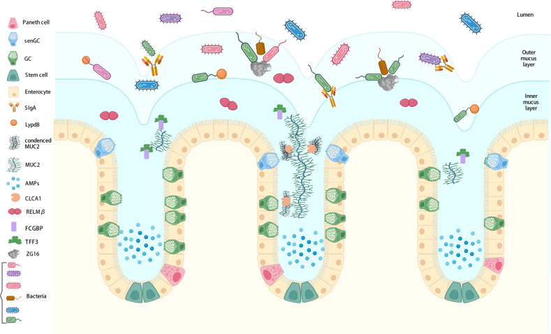
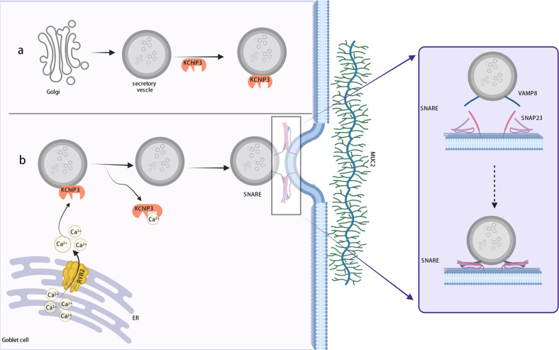
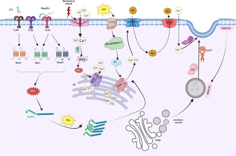
Damage to the colon mucus barrier, the first line of defense against microorganisms, is an important determinant of intestinal diseases such as inflammatory bowel disease and colorectal cancer, and disorder in extraintestinal organs. The mucus layer has attracted the attention of the scientific community in recent years, and with the discovery of new mucosal components, it has become increasingly clear that the mucosal barrier is a complex system composed of many components. Moreover, certain components are jointly involved in regulating the structure and function of the mucus barrier. Therefore, a comprehensive and systematic understanding of the functional components of the mucus layer is clearly warranted. In this review, we summarize the various functional components of the mucus layer identified thus far and describe their unique roles in shaping mucosal structure and function. Furthermore, we detail the mechanisms underlying mucus secretion, including baseline and stimulated secretion. In our opinion, baseline secretion can be categorized into spontaneous Ca2+ oscillation-mediated slow and continuous secretion and stimulated secretion, which is mediated by massive Ca2+ influx induced by exogenous stimuli. This review extends the current understanding of the intestinal mucus barrier, with an emphasis on host defense strategies based on fortification of the mucus layer.
© 2023. The Author(s).
Conflict of interest statement
The authors declare no competing interests.
Figures
Similar articles
-
Development and Functional Properties of Intestinal Mucus Layer in Poultry.Front Immunol. 2021 Oct 4;12:745849. doi: 10.3389/fimmu.2021.745849. eCollection 2021. Front Immunol. 2021. PMID: 34671361 Free PMC article. Review.
-
Gut Microbiota and Dietary Factors as Modulators of the Mucus Layer in Inflammatory Bowel Disease.Int J Mol Sci. 2021 Sep 23;22(19):10224. doi: 10.3390/ijms221910224. Int J Mol Sci. 2021. PMID: 34638564 Free PMC article. Review.
-
[Research progress of intestinal mucus barrier and the repair of intestinal damage after burn injury].Zhonghua Shao Shang Yu Chuang Mian Xiu Fu Za Zhi. 2022 Aug 20;38(8):788-793. doi: 10.3760/cma.j.cn501225-20220701-00280. Zhonghua Shao Shang Yu Chuang Mian Xiu Fu Za Zhi. 2022. PMID: 36058702 Free PMC article. Review. Chinese.
-
Gastroduodenal mucus bicarbonate barrier: protection against acid and pepsin.Am J Physiol Cell Physiol. 2005 Jan;288(1):C1-19. doi: 10.1152/ajpcell.00102.2004. Am J Physiol Cell Physiol. 2005. PMID: 15591243 Review.
-
Intestinal mucus barrier in normal and inflamed colon.J Pediatr Gastroenterol Nutr. 2009 Apr;48 Suppl 2:S54-5. doi: 10.1097/MPG.0b013e3181a117ea. J Pediatr Gastroenterol Nutr. 2009. PMID: 19300126
Cited by
-
Potent Intestinal Mucosal Barrier Enhancement of Nostoc commune Vaucher Polysaccharide Supplementation Ameliorates Acute Ulcerative Colitis in Mice Mediated by Gut Microbiota.Nutrients. 2023 Jul 6;15(13):3054. doi: 10.3390/nu15133054. Nutrients. 2023. PMID: 37447380 Free PMC article.
-
Mucin levels in glands of the colonic mucosa of rats with diversion colitis subjected to enemas containing sucralfate and n-acetylcysteine alone or in combination.Acta Cir Bras. 2023 Oct 13;38:e384023. doi: 10.1590/acb384023. eCollection 2023. Acta Cir Bras. 2023. PMID: 37851785 Free PMC article.
-
Overcoming Challenges in Small-Molecule Drug Bioavailability: A Review of Key Factors and Approaches.Int J Mol Sci. 2024 Dec 6;25(23):13121. doi: 10.3390/ijms252313121. Int J Mol Sci. 2024. PMID: 39684832 Free PMC article. Review.
-
Long-term Pu-erh tea alleviates inflammatory bowel disease via the regulation of intestinal microbiota and maintaining the intestinal mucosal barrier.Food Sci Biotechnol. 2024 Sep 2;34(3):743-755. doi: 10.1007/s10068-024-01696-9. eCollection 2025 Feb. Food Sci Biotechnol. 2024. PMID: 39958166
-
From Dysbiosis to Hepatic Inflammation: A Narrative Review on the Diet-Microbiota-Liver Axis in Steatotic Liver Disease.Microorganisms. 2025 Jan 23;13(2):241. doi: 10.3390/microorganisms13020241. Microorganisms. 2025. PMID: 40005608 Free PMC article. Review.
References
Publication types
MeSH terms
LinkOut - more resources
Full Text Sources
Miscellaneous
