. 2023 Apr;55(4):831-843.
doi: 10.1038/s12276-023-00972-8. Epub 2023 Apr 3.

LncRNA KIAA0087 suppresses the progression of osteosarcoma by mediating the SOCS1/JAK2/STAT3 signaling pathway

Affiliations

LncRNA KIAA0087 suppresses the progression of osteosarcoma by mediating the SOCS1/JAK2/STAT3 signaling pathway

Haoli Gong et al. Exp Mol Med. 2023 Apr.

Abstract

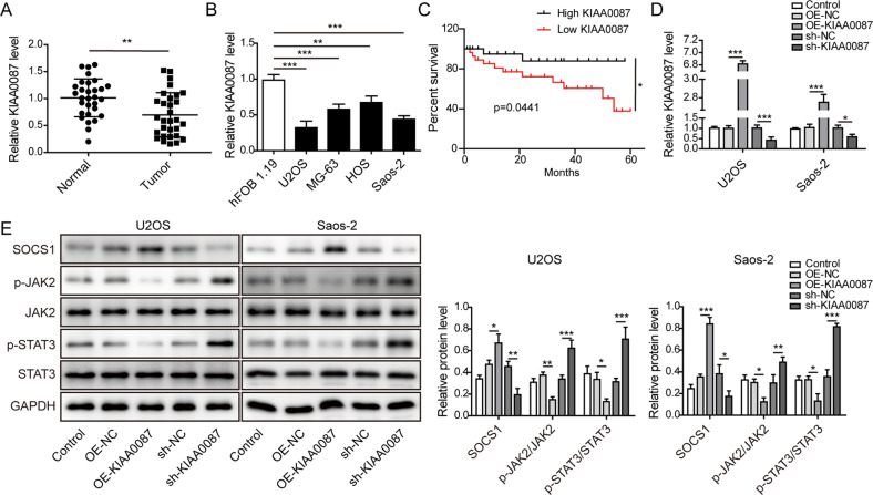
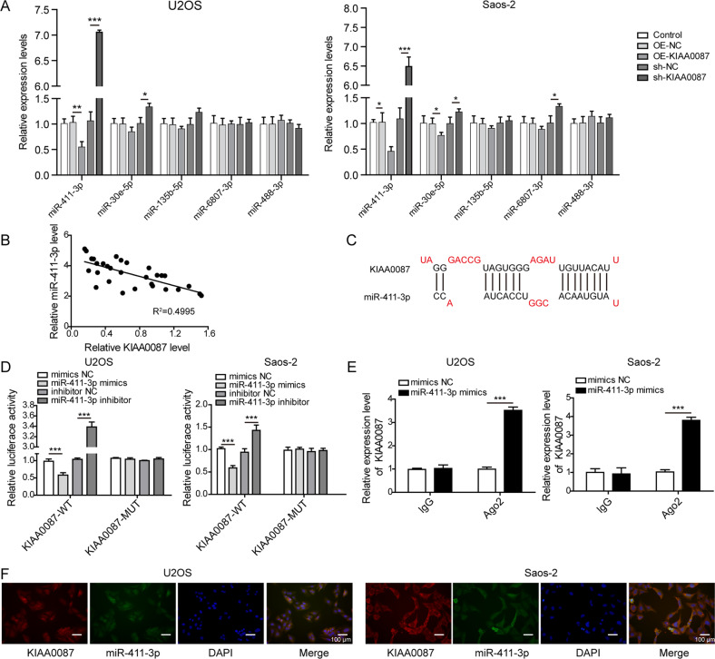
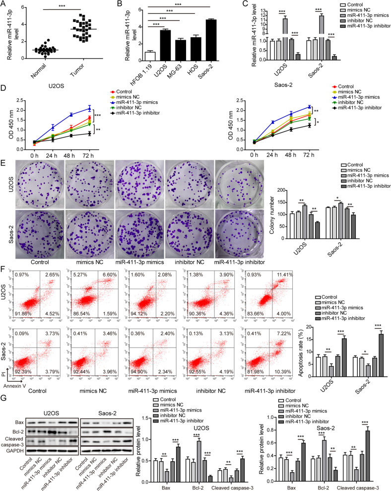
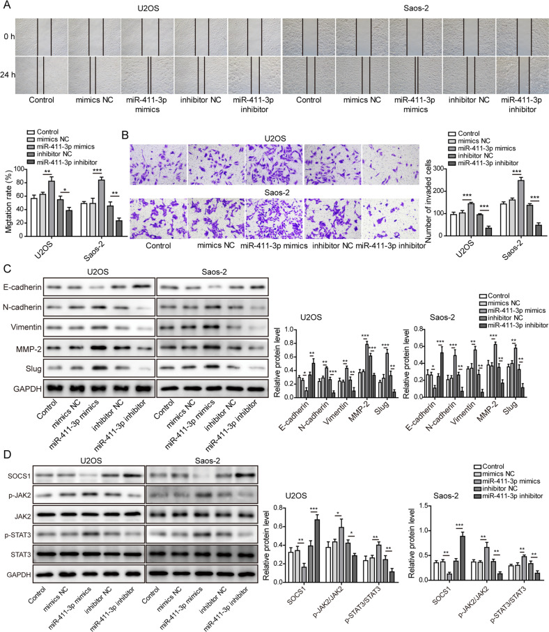
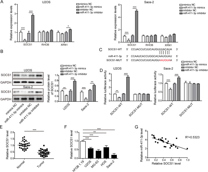
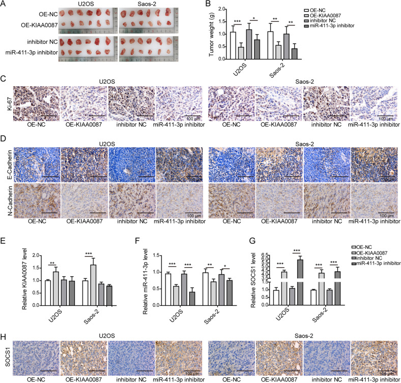
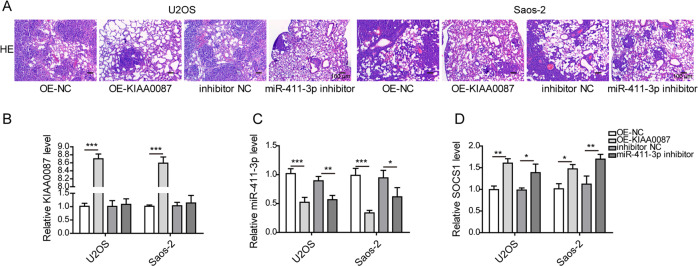
Long noncoding RNAs (lncRNAs), widely expressed in mammalian cells, play pivotal roles in osteosarcoma (OS) progression. Nevertheless, the detailed molecular mechanisms of lncRNA KIAA0087 in OS remain obscure. Here, the roles of KIAA0087 in OS tumorigenesis were investigated. KIAA0087 and miR-411-3p levels were detected by RT-qPCR. Malignant properties were assessed by CCK-8, colony formation, flow cytometry, wound healing, and transwell assays. SOCS1, EMT, and JAK2/STAT3 pathway-related protein levels were measured by western blotting. Direct binding between miR-411-3p and KIAA0087/SOCS1 was validated by a dual-luciferase reporter, RIP, and FISH assays. In vivo growth and lung metastasis were evaluated in nude mice. The expression levels of SOCS1, Ki-67, E-cadherin, and N-cadherin in tumor tissues were measured by immunohistochemical staining. Downregulation of KIAA0087 and SOCS1 and upregulation of miR-411-3p were found in OS tissues and cells. Low expression of KIAA0087 was associated with a poor survival rate. Forced expression of KIAA0087 or miR-411-3p inhibition repressed the growth, migration, invasion, EMT, and activation of the JAK2/STAT3 pathway and triggered apoptosis of OS cells. However, the opposite results were found with KIAA0087 knockdown or miR-411-3p overexpression. Mechanistic experiments indicated that KIAA0087 enhanced SOCS1 expression to inactivate the JAK2/STAT3 pathway by sponging miR-411-3p. Rescue experiments revealed that the antitumor effects of KIAA0087 overexpression or miR-411-3p suppression were counteracted by miR-411-3p mimics or SOCS1 inhibition, respectively. Finally, in vivo tumor growth and lung metastasis were inhibited in KIAA0087-overexpressing or miR-411-3p-inhibited OS cells. In summary, the downregulation of KIAA0087 promotes the growth, metastasis, and EMT of OS by targeting the miR-411-3p-mediated SOCS1/JAK2/STAT3 pathway.


Conflict of interest statement

The authors declare no competing interests.

Figures

Fig. 1
Fig. 1. Aberrant low expression of KIAA0087 in OS is associated with a poor prognosis.
a RT-qPCR analysis was used to evaluate KIAA0087 levels in clinically normal and OS samples (n = 30). b RT-qPCR analysis was used to evaluate KIAA0087 levels in hFOB 1.19 and different OS cells (U2OS, Saos-2, MG-63, and HOS). c The survival rate of OS patients with high or low expression of KIAA0087. d The transfection efficiency of overexpression or silencing of KIAA0087 was detected by RT-qPCR. e Western blot analysis of the protein levels of SOCS1, p-JAK2, JAK2, p-STAT3, and STAT3. *p < 0.05, **p < 0.01, and ***p < 0.001.
Fig. 2
Fig. 2. Dysregulation of KIAA0087 affects the proliferation and apoptosis of OS cells.
a CCK-8 assay determined the proliferation of OS cells. b Colony formation assay for evaluating the growth of OS cells. c Flow cytometry for testing the apoptotic rate of OS cells. d The protein levels of Bax, bcl-2, and cleaved caspase-3 in OS cells were determined by western blotting. *p < 0.05, **p < 0.01, and ***p < 0.001.
Fig. 3
Fig. 3. Effect of KIAA0087 on the migration, invasion, and EMT of OS cells.
a Wound healing assay for detecting the migration of OS cells. b Transwell assay for assessing the invasion of OS cells. c The protein levels of E-cadherin, N-cadherin, vimentin, MMP-2, and slug were assessed by western blotting. *p < 0.05, **p < 0.01, and ***p < 0.001.
Fig. 4
Fig. 4. KIAA0087 directly binds to miR-411-3p in OS cells.
a RT-qPCR detected miR-411-3p, miR-30e-5p, miR-135b-5p, miR-6807-3p, and miR-488-3p expression after overexpression or inhibition of KIAA0087 in OS cells. b The correlation between KIAA0087 and miR-411-3p expression in the clinical OS samples was evaluated (n = 30). c Bioinformatic analysis of the specific binding regions between KIAA0087 and miR-411-3p. d, e The direct binding of KIAA0087 to miR-411-3p was validated by dual-luciferase reporter assay (d) and RIP assay (e). f The cytoplasmic colocalization of KIAA0087 and miR-411-3p in OS cells was observed by FISH. *p < 0.05, **p < 0.01, and ***p < 0.001.
Fig. 5
Fig. 5. Upregulation of miR-411-3p confers OS cell malignant growth.
a RT-qPCR analysis was used to evaluate miR-411-3p levels in clinically normal and OS samples (n = 30). b RT-qPCR analysis was used to evaluate miR-411-3p levels in hFOB 1.19 and different OS cells (U2OS, Saos-2, MG-63, and HOS). c RT-qPCR assay was used to validate lentivirus-mediated overexpression or inhibition of miR-411-3p in OS cells. d CCK-8 assay determined the proliferation of OS cells. e Colony formation assay for evaluating the growth of OS cells. f Flow cytometry for testing the apoptotic rate of OS cells. g Western blot analysis of the protein levels of Bax, bcl-2, and cleaved caspase-3 in OS cells. *p < 0.05, **p < 0.01, and ***p < 0.001.
Fig. 6
Fig. 6. MiR-411-3p overexpression promotes OS cell migration, invasion, and EMT.
a Wound healing assay for detecting migration in OS cells. b Transwell assay for assessing the invasion of OS cells. c Western blotting for assessing the levels of EMT-related proteins, including E-cadherin, N-cadherin, vimentin, MMP-2, and slug. d Western blot analysis of the expression of SOCS1 and JAK2/STAT3 pathway component proteins. *p < 0.05, **p < 0.01, and ***p < 0.001.
Fig. 7
Fig. 7. SOCS1 is a target gene of miR-411-3p in OS cells.
a The mRNA levels of SOCS1, RHOB, and XRN1 were detected by RT-qPCR after overexpression or inhibition of miR-411-3p in OS cells. b Western blotting was used to evaluate the protein level of SOCS1 in miR-411-3p mimic- or inhibitor-transfected OS cells. c Bioinformatic analysis of the potential miR-411-3p binding sites within the SOCS1 3′-UTR. d The direct binding of miR-411-3p to SOCS1 was confirmed by dual-luciferase reporter assay. e The expression level of SOCS1 in clinically normal and OS samples (n = 30) was determined by RT-qPCR. f RT-qPCR analysis of SOCS1 mRNA levels in various OS cells (U2OS, Saos-2, MG-63, and HOS). g Spearman correlation analysis of the correlation between the expression of miR-411-3p and SOCS1 in clinical OS samples (n = 30). *p < 0.05, **p < 0.01, and ***p < 0.001.
Fig. 8
Fig. 8. KIAA0087 overexpression or miR-411-3p knockdown disrupts tumor growth in vivo.
a, b Xenograft tumors (a) and quantitative results (b) of tumor weight from various groups. c Ki-67 expression in tumor tissues was determined by immunohistochemical staining. d E-cadherin and N-cadherin expression in tumor tissues was determined by immunohistochemical staining. eg RT-qPCR for determining KIAA0087 (e), miR-411-3p (f), and SOCS1 (g) expression in tumor tissues. h SOCS1 expression in tumor tissues was detected by immunohistochemical staining. *p < 0.05, **p < 0.01, and ***p < 0.001.
Fig. 9
Fig. 9. KIAA0087 overexpression or miR-411-3p inhibition suppresses lung metastasis in vivo.
a HE staining for evaluating metastatic tumor cells in lung tissues. bd RT-qPCR for detecting KIAA0087 (b), miR-411-3p (c), and SOCS1 (d) expression in the lung tissues of nude mice. *p < 0.05, **p < 0.01, and ***p < 0.001.

References

    1. Caudill JS, Arndt CA. Diagnosis and management of bone malignancy in adolescence. Adolesc. Med. State Art. Rev. 2007;18:62–78. - PubMed
    1. Ottaviani G, Jaffe N. The epidemiology of osteosarcoma. Cancer Treat. Res. 2009;152:3–13. doi: 10.1007/978-1-4419-0284-9_1. - DOI - PubMed
    1. Tsukamoto S, Errani C, Angelini A, Mavrogenis AF. Current treatment considerations for osteosarcoma metastatic at presentation. Orthopedics. 2020;43:e345–e358. doi: 10.3928/01477447-20200721-05. - DOI - PubMed
    1. Qiu S, Tao L, Zhu Y. Marital status and survival in osteosarcoma patients: an analysis of the surveillance, epidemiology, and end results (SEER) database. Med. Sci. Monit. 2019;25:8190–8203. doi: 10.12659/MSM.918048. - DOI - PMC - PubMed
    1. Lu KH. Molecular and cellular mechanisms of melatonin in osteosarcoma. Cells. 2019;8:1618. doi: 10.3390/cells8121618. - DOI - PMC - PubMed

Publication types