. 2023 Apr 3;133(7):e161913.
doi: 10.1172/JCI161913.

Cancer-associated fibroblast-secreted glucosamine alters the androgen biosynthesis program in prostate cancer via HSD3B1 upregulation

Affiliations

Cancer-associated fibroblast-secreted glucosamine alters the androgen biosynthesis program in prostate cancer via HSD3B1 upregulation

Di Cui et al. J Clin Invest. .

Abstract

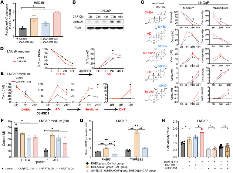
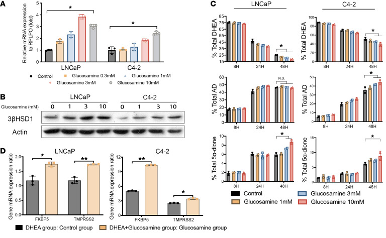
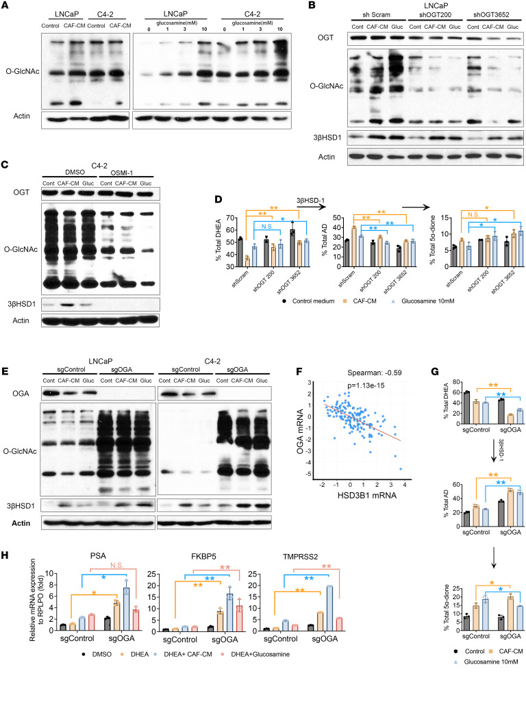
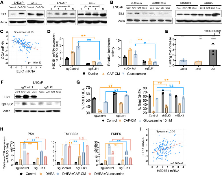
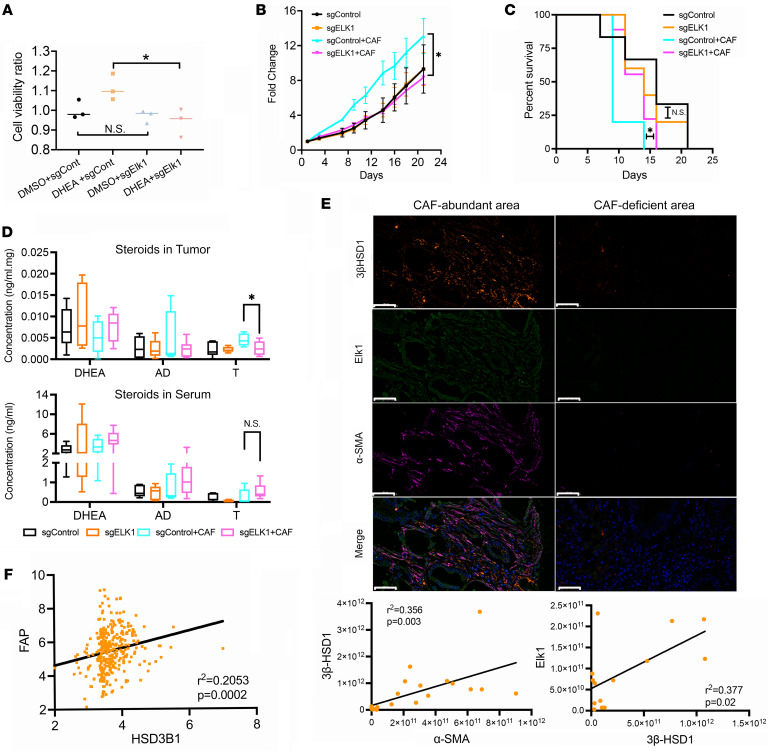
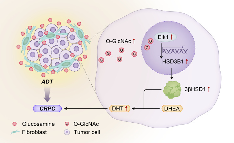
After androgen deprivation, prostate cancer frequently becomes castration resistant (CRPC), with intratumoral androgen production from extragonadal precursors that activate the androgen receptor pathway. 3β-Hydroxysteroid dehydrogenase-1 (3βHSD1) is the rate-limiting enzyme for extragonadal androgen synthesis, which together lead to CRPC. Here, we show that cancer-associated fibroblasts (CAFs) increased epithelial 3βHSD1 expression, induced androgen synthesis, activated the androgen receptor, and induced CRPC. Unbiased metabolomics revealed that CAF-secreted glucosamine specifically induced 3βHSD1. CAFs induced higher GlcNAcylation in cancer cells and elevated expression of the transcription factor Elk1, which induced higher 3βHSD1 expression and activity. Elk1 genetic ablation in cancer epithelial cells suppressed CAF-induced androgen biosynthesis in vivo. In patient samples, multiplex fluorescent imaging showed that tumor cells expressed more 3βHSD1 and Elk1 in CAF-enriched areas compared with CAF-deficient areas. Our findings suggest that CAF-secreted glucosamine increases GlcNAcylation in prostate cancer cells, promoting Elk1-induced HSD3B1 transcription, which upregulates de novo intratumoral androgen synthesis to overcome castration.

Keywords: Oncology; Prostate cancer; Sex hormones.


Conflict of interest statement

Conflict of interest: NS is a coinventor on Cleveland Clinic US patent US20180023146A1, 3-β-hydroxysteroid dehydrogenase in steroid-dependent disease. NS has received research funding from BMS and Astellas.

Figures

Figure 1
Figure 1. CAFs increase the conversion from DHEA to active androgens in LNCaP cells by increasing 3βHSD1 expression and enzyme activity.
(A) mRNA (by qPCR) and (B) protein expression of HSD3B1 and 3βHSD1 in LNCaP cells treated with CAF–conditioned medium (CAF-CM) for the indicated times. Gene and protein expression was normalized to RPLP0 and β-actin, respectively. (C) LNCaP cells were treated with CAF-CM for 48 hours followed by 100 nM DHEA for the indicated times. Downstream androgens in intracellular and media samples were quantitated by mass spectrometry. (D) LNCaP cells were treated with CAF-CM for 48 hours, followed by [3H]-DHEA (106 counts per minute) for the indicated times, followed by extraction of steroids from medium and quantitation by HPLC. (E) Mass spectrometry analysis of steroids in medium of LNCaP cells treated with DHEA along with CAF-CM derived from primary CAFs isolated from fresh prostate tumor tissue (patient no. 1). (F) Mass spectrometry analysis of primary CAF-CM from 3 patients with prostate cancer (patients 2, 3, and 4). (G) AR target gene (FKBP5 and TMPRSS2) mRNA expression in LNCaP cells (control or HSD3B1 siRNA; treated with 10nM DHEA and CAF-CM for 48 hours). Expression was normalized to untreated cells (data not shown), and RPLP0 was used as a loading control. (H) Cell viability of LNCaP control or HSD3B1 siRNA cells treated with DHEA along with control media or CAF-CM. Viability was normalized to the untreated control. Unless otherwise noted, data are shown as mean ± SEM. Significance was calculated using 2-tailed t tests or 1-way ANOVA as appropriate. *P < 0.05, **P < 0.01. Conc., Concentration.
Figure 2
Figure 2. Glucosamine in CAF-CM induces HSD3B1 expression and the androgen metabolism phenotype.
(A) mRNA (qPCR) and (B) protein expression of 3βHSD1 in LNCaP and C4-2 cells treated with increasing concentrations of glucosamine for 48 hours. Significance was calculated using 2-tailed t tests (control versus 10 mM glucosamine). (C) HPLC analysis of steroids in media of C4-2 and LNCaP cells treated with the indicated concentrations of glucosamine for 48 hours and [3H]-DHEA (1,000,000 counts per minute) for the indicated times. Significance was calculated at 48 hours using 1-way ANOVA. (D) Gene expression of AR target genes in C42 and LNCaP cells treated with 10 nM DHEA in the presence or absence of 10 mM glucosamine. Data were normalized to RPLP0, and significance was calculated using 2-tailed t tests. *P < 0.05.
Figure 3
Figure 3. O-GlcNAcylation after CAF-CM treatment is attributable to glucosamine and induces 3βHSD1 expression and enzyme activity.
(A) Western blot analysis of O-GlcNAcylated proteins in LNCaP and C42 cells treated with CAF–conditioned medium (CAF-CM) (left) or increasing concentrations of glucosamine (right) for 48 hours. (B) Western blot analysis of O-GlcNAcylated proteins, OGT, and 3βHSD1 protein expression in LNCaP cells expressing 2 shRNAs targeting OGT (OCG200 and OGT3652) or scrambled shRNA and treated with CAF-CM or 10 mM glucosamine for 48 hours. (C) O-GlcNAcylated proteins, OGT, and 3βHSD1 protein expression in C4-2 cells treated with CAF-CM or 10 mM glucosamine in the presence of DMSO (vehicle control) or 75 μM OSMI-1, an OGT inhibitor. (D) HPLC analysis of DHEA metabolism in LNCaP cells expressing scrambled shRNA, shOCG200, and shOGT3652 treated with CAF-CM or 10 mM glucosamine for 48 hours, followed by addition of [3H]-DHEA (100 nM) for the indicated times. Significance was calculated using 1-way ANOVA. (E) Western blot analysis of OGA, O-GlcNAcylated proteins, and 3βHSD1 expression in LNCaP (left) and C4-2 (right) cells transduced with control (sgControl) or sgRNA targeting OGA (sgOGA) after 48-hour treatment with CAF-CM or 10 mM glucosamine. (F) Pearson correlation analysis of HSD3B1 and OGA mRNA expression in prostate cancer (MSKCC Prostate Oncogenome Project, GSE21032). (G) HPLC analysis of DHEA metabolism in media of LNCaP control or OGA-KO cells treated with CAF-CM or glucosamine for 48 hours, followed by [3H]-DHEA (100 nM) for the indicated times; data were normalized to untreated control. (H) Gene expression of AR target genes PSA, FKBP5, and TMPRSS2 in control and OGA-KO LNCaP cells treated with CAF-CM or 10 mM glucosamine plus 10 nM DHEA for 48 hours. Significance was calculated using 2-tailed t tests. *P < 0.05, **P < 0.01.
Figure 4
Figure 4. High O-GlcNAcylation increases Elk1 to induce 3βHSD1 expression and enzyme activity.
(A) Protein expression of ELK1 in LNCaP and C42 cells treated with CAF–conditioned medium (CAF-CM) (left) and increasing concentrations of glucosamine (right) for 48 hours. (B) ELK1 protein expression in LNCaP cells expressing control or OGT shRNA (left) and sgRNA (right) treated with CAF-CM or 10 mM glucosamine for 48 hours. (C) Pearson correlation analysis of ELK1 and OGA mRNA expression in prostate cancer (GSE21032). (D) Left: Gene expression of HSD3B1 in control (sgControl) or ELK1-KO (sgELK1) LNCaP cells treated with CAF-CM or 10 mM glucosamine for 48 hours. Right: Luciferase assay of LNCaP control and ELK1-KO cells cotransfected with an HSD3B1 promoter-firefly luciferase and Renilla luciferase plasmid constructs, which were treated with CAF-CM or 10 mM glucosamine 48 hours. (E) ChIP assay of Elk1. C4-2 cells were treated with 10 mM glucosamine for 24 hours. (F) Protein expression of ELK1 and 3βHSD1 in sgControl and ELK1-KO LNCaP cells treated with CAF-CM or glucosamine. (G) HPLC analysis of steroids in media of LNCaP cells expressing sgControl and ELK1 KO (left) or siRNA and shRNA knockdown of ELK1 (right). Cells were treated with CAF-CM or glucosamine for 48 hours, followed by [3H]-DHEA (100 nM) for 48 hours. (H) Gene expression of PSA, TMPRSS2, and FKBP5 in sgControl and ELK1-KO LNCaP cells treated with 10 nM DHEA along with CAF-CM or glucosamine for 48 hours. (I) Pearson correlation analysis of ELK1 and HSD3B1 mRNA expression in prostate cancer (GSE21032). Significance was calculated using 2-tailed t tests. *P < 0.05, **P < 0.01.
Figure 5
Figure 5. Elk1 induced 3βHSD1 expression and DHEA metabolism in vivo.
(A) Cell viability of sgControl and ELK1-KO LNCaP cells treated with DMSO (control) or 10 nM DHEA for 48 hours. (B) Xenograft tumor growth of orchiectomized mice subcutaneously injected with control or ELK1-KO C42 cells in the absence or presence of CAFs. A 2-tailed paired t test was performed between control and ELK1-KO tumors coinjected with CAFs at day 21. (C) A log-rank test was used to compare progression-free survival between control and ELK1-KO and C4-2 cells grown with CAFs. (D) Mass spectrometry analysis of intratumoral and serum DHEA, AD, and testosterone (T) in control or ELK1-KO C42 cells. (E) Representative multiplexed fluorescence image of a patient with prostate cancer (Gleason 4+4). 3βHSD1, orange; Elk1, green; CAF, α-SMA, purple), and DAPI, blue. Scale bar: 50 μm. Pearson correlation analysis of gene expression in tissues from patients with primary prostate cancer (3βHSD1 and CAF [α-SMA], n = 22; 3βHSD1 and Elk1, n = 14). (F) Pearson correlation analysis of HSD3B1 and FAP mRNA (CAF) in human CRPC metastases (GSE77930). Unless otherwise noted, data are shown as mean ± SEM. Significance was calculated using a 2-tailed t test or 1-way ANOVA. *P < 0.05, **P < 0.01.
Figure 6
Figure 6. The physiology and mechanisms by which glucosamine originating from fibroblasts induces androgen biosynthesis and resistance in prostate cancer.
Cancer-associated fibroblasts synthesize and secrete glucosamine in the tumor microenvironment. In the prostate cancer tumor cell, glucosamine induces an increase in O-GlcNAc, which in turn elicits Elk1-dependent transcription of HSD3B1. HSD3B1 is translated to its corresponding enzyme, 3βHSD1, which is the rate-limiting step for prostate cancer to convert adrenal DHEA to the potent androgen, DHT, to promote progression to castration-resistant prostate cancer (CRPC).

Comment in

  • CAFs promote CRPC.
    Brewer G. Brewer G. Nat Rev Cancer. 2023 Jun;23(6):349. doi: 10.1038/s41568-023-00587-1. Nat Rev Cancer. 2023. PMID: 37173415 No abstract available.
  • Uro-Science.
    Atala A. Atala A. J Urol. 2023 Dec;210(6):922-924. doi: 10.1097/JU.0000000000003662. Epub 2023 Sep 29. J Urol. 2023. PMID: 37774378 No abstract available.

Similar articles

Cited by

References

    1. Desai K, et al. Hormonal therapy for prostate cancer. Endocr Rev. 2021;42(3):354–373. doi: 10.1210/endrev/bnab002. - DOI - PMC - PubMed
    1. Dai C, et al. Androgen signaling in prostate cancer. Cold Spring Harb Perspect Med. 2017;7(9):a030452. doi: 10.1101/cshperspect.a030452. - DOI - PMC - PubMed
    1. Mostaghel EA, et al. Circulating and intratumoral adrenal androgens correlate with response to abiraterone in men with castration-resistant prostate cancer. Clin Cancer Res. 2021;27(21):6001–6011. doi: 10.1158/1078-0432.CCR-21-1819. - DOI - PMC - PubMed
    1. Davies AH, Zoubeidi A. Targeting androgen receptor signaling: a historical perspective. Endocr Relat Cancer. 2021;28(8):T11–T18. doi: 10.1530/ERC-21-0116. - DOI - PubMed
    1. Watson PA, et al. Emerging mechanisms of resistance to androgen receptor inhibitors in prostate cancer. Nat Rev Cancer. 2015;15(12):701–711. doi: 10.1038/nrc4016. - DOI - PMC - PubMed

Publication types