

Need for objective task-based evaluation of deep learning-based denoising methods: A study in the context of myocardial perfusion SPECT
- PMID: 37010001
- PMCID: PMC10524194
- DOI: 10.1002/mp.16407
Need for objective task-based evaluation of deep learning-based denoising methods: A study in the context of myocardial perfusion SPECT
Abstract
Background: Artificial intelligence-based methods have generated substantial interest in nuclear medicine. An area of significant interest has been the use of deep-learning (DL)-based approaches for denoising images acquired with lower doses, shorter acquisition times, or both. Objective evaluation of these approaches is essential for clinical application.
Purpose: DL-based approaches for denoising nuclear-medicine images have typically been evaluated using fidelity-based figures of merit (FoMs) such as root mean squared error (RMSE) and structural similarity index measure (SSIM). However, these images are acquired for clinical tasks and thus should be evaluated based on their performance in these tasks. Our objectives were to: (1) investigate whether evaluation with these FoMs is consistent with objective clinical-task-based evaluation; (2) provide a theoretical analysis for determining the impact of denoising on signal-detection tasks; and (3) demonstrate the utility of virtual imaging trials (VITs) to evaluate DL-based methods.
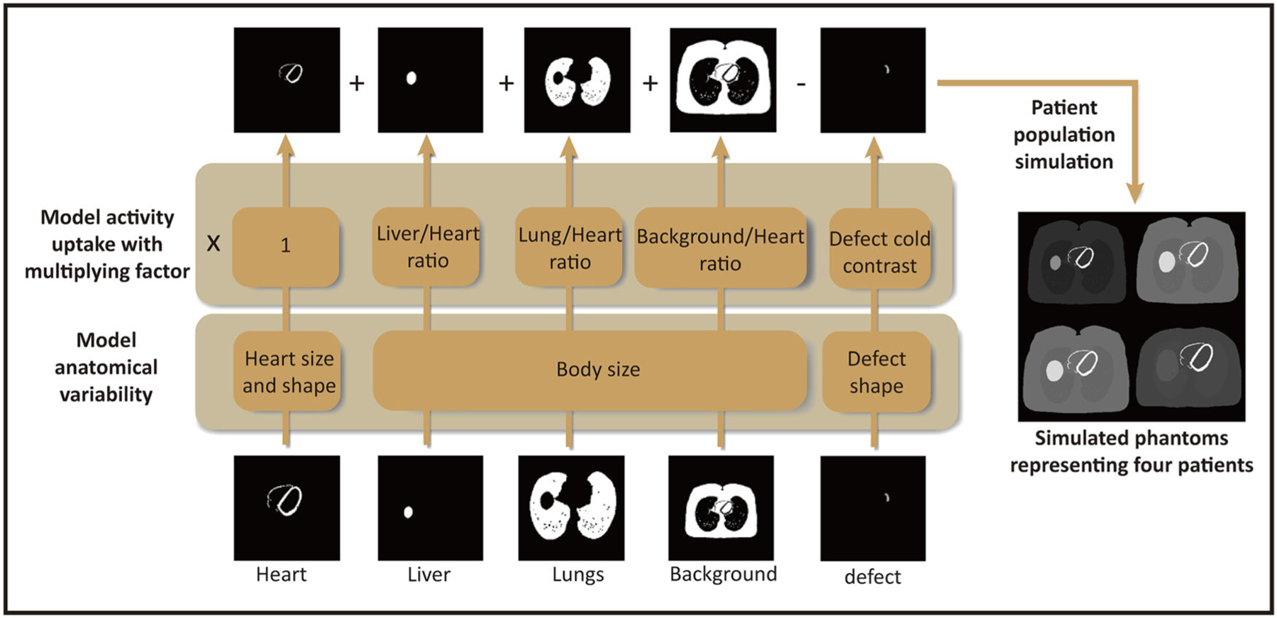
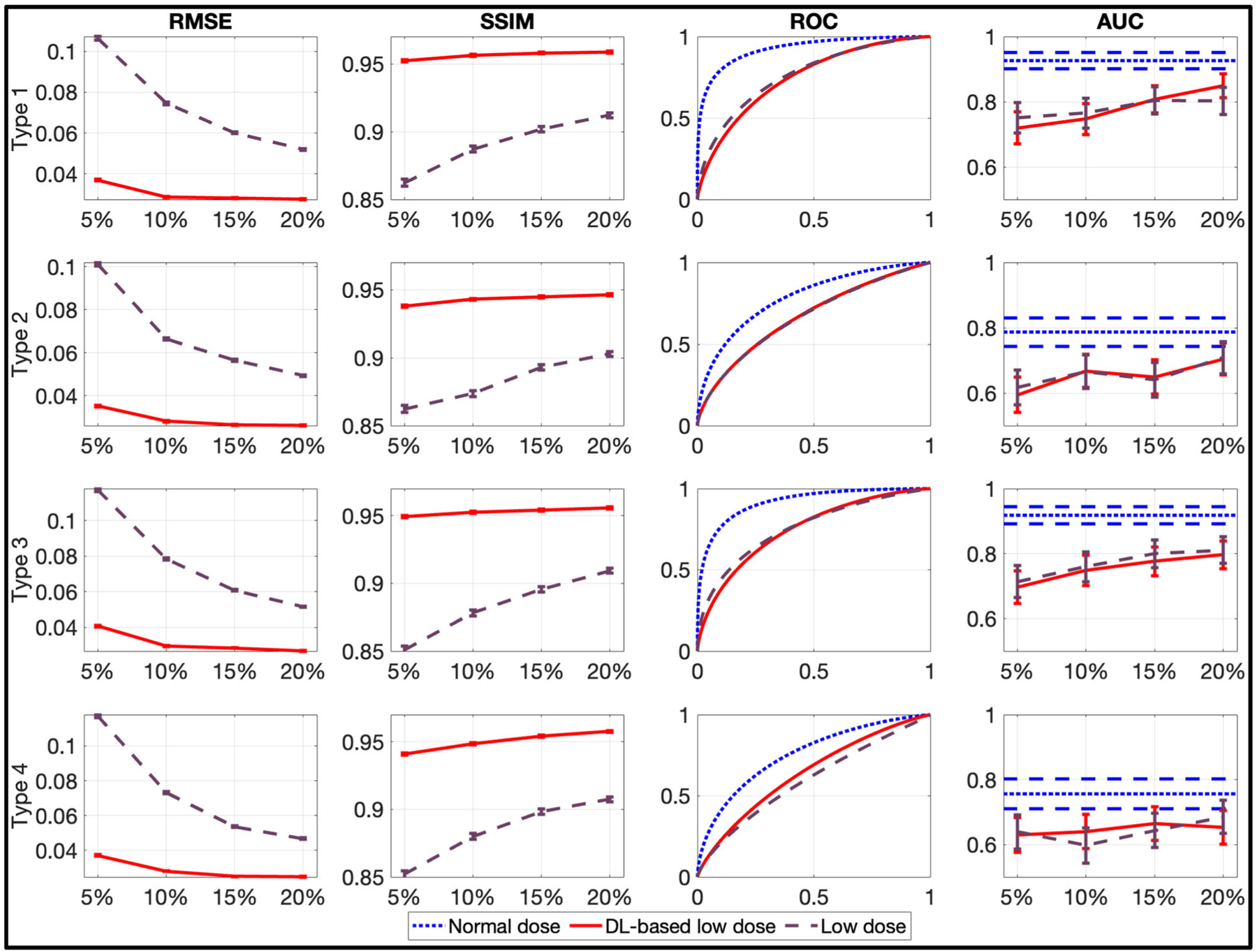
Methods: A VIT to evaluate a DL-based method for denoising myocardial perfusion SPECT (MPS) images was conducted. To conduct this evaluation study, we followed the recently published best practices for the evaluation of AI algorithms for nuclear medicine (the RELAINCE guidelines). An anthropomorphic patient population modeling clinically relevant variability was simulated. Projection data for this patient population at normal and low-dose count levels (20%, 15%, 10%, 5%) were generated using well-validated Monte Carlo-based simulations. The images were reconstructed using a 3-D ordered-subsets expectation maximization-based approach. Next, the low-dose images were denoised using a commonly used convolutional neural network-based approach. The impact of DL-based denoising was evaluated using both fidelity-based FoMs and area under the receiver operating characteristic curve (AUC), which quantified performance on the clinical task of detecting perfusion defects in MPS images as obtained using a model observer with anthropomorphic channels. We then provide a mathematical treatment to probe the impact of post-processing operations on signal-detection tasks and use this treatment to analyze the findings of this study.
Results: Based on fidelity-based FoMs, denoising using the considered DL-based method led to significantly superior performance. However, based on ROC analysis, denoising did not improve, and in fact, often degraded detection-task performance. This discordance between fidelity-based FoMs and task-based evaluation was observed at all the low-dose levels and for different cardiac-defect types. Our theoretical analysis revealed that the major reason for this degraded performance was that the denoising method reduced the difference in the means of the reconstructed images and of the channel operator-extracted feature vectors between the defect-absent and defect-present cases.
Conclusions: The results show the discrepancy between the evaluation of DL-based methods with fidelity-based metrics versus the evaluation on clinical tasks. This motivates the need for objective task-based evaluation of DL-based denoising approaches. Further, this study shows how VITs provide a mechanism to conduct such evaluations computationally, in a time and resource-efficient setting, and avoid risks such as radiation dose to the patient. Finally, our theoretical treatment reveals insights into the reasons for the limited performance of the denoising approach and may be used to probe the effect of other post-processing operations on signal-detection tasks.
Keywords: SPECT; deep learning; model observer; task-based evaluation.
© 2023 American Association of Physicists in Medicine.
Conflict of interest statement
CONFLICT OF INTEREST STATEMENT
No potential conflicts of interest relevant to this article exist.
Figures


Update of
-
Need for Objective Task-based Evaluation of Deep Learning-Based Denoising Methods: A Study in the Context of Myocardial Perfusion SPECT.ArXiv [Preprint]. 2023 Apr 2:arXiv:2303.02110v5. ArXiv. 2023. Update in: Med Phys. 2023 Jul;50(7):4122-4137. doi: 10.1002/mp.16407. PMID: 36945690 Free PMC article. Updated. Preprint.
References
-
- Barrett HH. Objective assessment of image quality - effects of quantum noise and object variability. J Opt Soc Am A: Opt Image Sci Vis 1990;7(7):1266–1278. - PubMed
-
- Barrett HH, Denny JL, Wagner RF, Myers KJ. Objective assessment of image quality. II. Fisher information, Fourier crosstalk, and figures of merit for task-performance. J Opt Soc Am A: Opt Image Sci Vis 1995;12(5):834–852. - PubMed
-
- Barrett HH, Abbey CK, Clarkson E. Objective assessment of image quality. III. ROC metrics, ideal observers, and likelihood-generating functions. J Opt Soc Am A: Opt Image Sci Vis 1998;15(6):1520–1535. - PubMed
-
- Frey EC, Gilland KL, Tsui BMW. Application of task-based measures of image quality to optimization and evaluation of three-dimensional reconstruction-based compensation methods in myocardial perfusion SPECT. IEEE Trans Med Imaging 2002;21(9):1040–1050. - PubMed
-
- He X, Frey EC, Links JM, Gilland KL, Segars WP, Tsui BMW. A mathematical observer study for the evaluation and optimization of compensation methods for myocardial SPECT using a phantom population that realistically models patient variability. IEEE Trans Nucl Sci 2004;51(1):218–224.
MeSH terms
Grants and funding
LinkOut - more resources
Full Text Sources
