. 2023 Jun 19;32(13):2177-2191.
doi: 10.1093/hmg/ddad054.

A humanized yeast model reveals dominant-negative properties of neuropathy-associated alanyl-tRNA synthetase mutations

Affiliations

A humanized yeast model reveals dominant-negative properties of neuropathy-associated alanyl-tRNA synthetase mutations

Rebecca Meyer-Schuman et al. Hum Mol Genet. .

Abstract

Aminoacyl-tRNA synthetases (ARSs) are essential enzymes that ligate tRNA molecules to cognate amino acids. Heterozygosity for missense variants or small in-frame deletions in six ARS genes causes dominant axonal peripheral neuropathy. These pathogenic variants reduce enzyme activity without significantly decreasing protein levels and reside in genes encoding homo-dimeric enzymes. These observations raise the possibility that neuropathy-associated ARS variants exert a dominant-negative effect, reducing overall ARS activity below a threshold required for peripheral nerve function. To test such variants for dominant-negative properties, we developed a humanized yeast assay to co-express pathogenic human alanyl-tRNA synthetase (AARS1) mutations with wild-type human AARS1. We show that multiple loss-of-function AARS1 mutations impair yeast growth through an interaction with wild-type AARS1, but that reducing this interaction rescues yeast growth. This suggests that neuropathy-associated AARS1 variants exert a dominant-negative effect, which supports a common, loss-of-function mechanism for ARS-mediated dominant peripheral neuropathy.


Conflict of interest statement

There is no conflict of interest to declare.

Figures

Figure 1
Figure 1
G102R and R329H AARS1 are loss-of-function alleles that repress yeast cell growth in the presence of wild-type AARS1. (A) Yeast harboring an endogenous doxycycline-repressible ALA1 locus were transformed with a p413 vector with no insert and a pAG425 vector to express either wild-type or mutant human AARS1. Cultures were plated undiluted or diluted on media lacking histidine and leucine, and containing galactose/raffinose and doxycycline. (B) Similar experiment as shown in panel A; here, yeast were first transformed with a p413 vector expressing wild-type human AARS1. For both panels, the vectors present in each experiment are indicated across the top, the dilution of the spotted yeast cultured is indicated on the left side of the image, and the media conditions are indicated across the bottom of the image (his = histidine; leu = leucine; gal = galactose; raf = raffinose; dox = doxycycline). Representative images are shown from thirteen (for G102R) or sixteen (for R329H) biological replicates. A cartoon on the bottom left illustrates the experimental conditions for all samples.
Figure 2
Figure 2
G102R and R329H AARS1 dimerize with wild-type AARS1. (A) HEK293T cells were transfected with vectors to co-express wild-type and mutant human AARS1, and a western blot was performed to detect the resulting proteins along with endogenous loading controls. The image is representative of three independent replicates. A cartoon along the top illustrates the constructs employed in the experiments, and the presence or absence of each construct is indicated across the top of the gel image. Protein molecular weights are indicated in kilodaltons (kDa) along the left side of the image and antibodies are indicated along the right side. (B) After immunoprecipitation with an anti-6xHis antibody, a western blot was performed to detect co-immunoprecipitated proteins. A representative image from five (for R329H) or three (for G102R) independent replicates are shown. This image is annotated as in panel A. (C) After immunoprecipitation with an anti-FLAG antibody, a western blot was performed to detect co-immunoprecipitated proteins. A representative image from two independent replicates is shown. This image is annotated as in panels A and B. (D) After treating patient and control samples with a protein cross-linking agent, a western blot was performed to detect endogenous AARS1 protein. The image is representative of four independent technical replicates. Samples and conditions are indicated across the top of the image, protein molecular weights are indicated in kilodaltons (kDa) along the left side, and the antibody employed is indicated on the right side. DSS = disuccinimidyl suberate. (E) The percentage of dimeric AARS1 protein signal in the total AARS1 protein signal (D) was quantified with ImageJ. The mean of four technical replicates is shown, with error bars representing one standard deviation. A one-way ANOVA with Tukey’s multiple comparisons tests [F(2,9) = 0.3602, P = 0.7071] was performed to determine if there was a statistically significant difference between R329H/+ cells and either of the two controls.
Figure 3
Figure 3
Engineering dimer-reducing AARS1 variants. (A) A cartoon generated from PyMOL illustrates the crystal structure of the AARS1 C-terminal dimerization domain (left side). One subunit from the dimer is shown in green, and the other in purple. Amino-acid residues that contact the opposite subunit are shown in dark green or dark purple. The residues targeted in this assay are shown in pink and labeled in the inset. On the right side of this panel is a second cartoon that illustrates the AARS1 C-terminal dimerization domain with the Q855* mutation. The dashed circles indicate the globular domain that is ablated by the premature stop codon. (B) Yeast harboring a doxycycline-repressible endogenous ALA1 locus were transformed with a pAG425 vector to express either wild-type or mutant human AARS1 (i.e. one of the engineered mutations affecting the residues highlighted in panel A). Cultures were plated undiluted or diluted on media lacking leucine, and containing galactose/raffinose and doxycycline. A representative image of four biological replicates is shown. The dilution of the spotted yeast cultured is indicated on the left and the media conditions are indicated across the bottom (leu = leucine; gal = galactose; raf = raffinose; dox = doxycycline). (C) Yeast protein lysates were subjected to western blot analysis to detect the human AARS1 proteins expressed from wild-type and mutant expression constructs, which are indicated across the top. Yeast was grown in galactose and raffinose media lacking leucine, with no doxycycline. A representative image of three biological replicates is shown.
Figure 4
Figure 4
Q855*AARS1 impairs dimerization with wild-type AARS1. (A) HEK293T cells were transfected with vectors to co-express wild-type or Q855* human AARS1, and a western blot was performed to detect the resulting proteins, as well as endogenous loading controls. The image is representative of three independent replicates. The presence or absence of each construct is shown across the top of the image, protein molecular weights are indicated in kilodaltons (kDa) along the left side, and antibodies are indicated along the right side. (B) After immunoprecipitation with an anti-6xHis antibody, a western blot was performed to detect co-immunoprecipitated proteins. The presence or absence of each construct is indicated at the top of the panel, and a representative image from three independent replicates is shown at the bottom of the panel (with annotation as in panel A). The middle of the panel shows ImageJ quantification of a 3xFLAG-tagged AARS1 signal. Bars indicate the mean value and one standard deviation for three biological replicates. A one-way ANOVA with Dunnett’s multiple comparisons test was performed to determine statistical significance [F(2,6) = 15.21, P = 0.0045]. (C) After immunoprecipitation with an anti-FLAG antibody, a western blot was performed to detect co-immunoprecipitated proteins. The panel is organized similarly to panel B, with ImageJ quantification of 6xHis-tagged AARS1 signal in the middle of the panel. Bars indicate the mean value and one standard deviation for three biological replicates. A one-way ANOVA with Dunnett’s multiple comparisons test was performed to determine statistical significance [F(2,6) = 191.7, P < 0.0001].
Figure 5
Figure 5
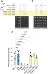
Reducing dimerization of G102R and R329H with wild-type AARS1 rescues yeast growth. (A) Yeast with the doxycycline-repressible endogenous ALA1 locus was transformed with an empty p413 vector and a pAG425 vector expressing wild-type or mutant human AARS1. Cultures were plated undiluted or diluted on media lacking histidine and leucine, and containing galactose/raffinose and doxycycline. (B) Similar to strains shown in panel A, except that yeast was transformed with p413 expressing wild-type human AARS1. For both panels, the vectors present in each experiment are indicated across the top, the dilution of the spotted yeast cultured is indicated on the left, and the media conditions are indicated across the bottom (his = histidine; leu = leucine; gal = galactose; raf = raffinose; dox = doxycycline). (C) Yeast spot intensity was quantified using ImageJ. Bars represent the mean and one standard deviation. Thirteen biological replicates were assessed for G757* and wild-type AARS1, eight for R329H and R329H + Q855*, and seven for G102R and G102R + Q855*. The indicated fold-change between the G102R strain and the G102R + Q855* strain, and the fold-change between the R329H strain and the R329H + Q855* strain, were both calculated using the mean of each sample. To compare yeast growth to the strain expressing both wild-type and G757*AARS1, a one-way ANOVA with Dunnett’s multiple comparisons test was performed [F(5,50) = 19.90, P < 0.0001].
Figure 6
Figure 6
R329C, R329S and R326W AARS1 are loss-of-function alleles that dominantly repress yeast cell growth. (A) Yeast with the doxycycline-repressible endogenous ALA1 locus was transformed with a p413 vector with no insert, and a pAG425 vector expressing wild-type or mutant AARS1. Yeast cultures were spotted undiluted or diluted on media lacking histidine and leucine, and containing galactose/raffinose and doxycycline. (B) A similar experiment to that described in panel A, except that yeast expresses wild-type human AARS1 from p413. For both panels, the vectors present in each experiment are indicated across the top, the dilution of the spotted yeast cultured is indicated on the left, and the media conditions are shown across the bottom (his = histidine; leu = leucine; gal = galactose; raf = raffinose; dox = doxycycline). Images are representative of three replicates.
Figure 7
Figure 7
Reducing dimerization of R329C, R329S or R326W with wild-type AARS1 rescues yeast growth. (A) Yeast with the doxycycline-repressible ALA1 locus was transformed with an empty p413 vector and a pAG425 vector expressing either wild-type or mutant AARS1. Yeast was spotted undiluted or diluted on media lacking histidine and leucine, and containing galactose/raffinose and doxycycline. (B) An experiment similar to that described in panel A, except that yeast expresses wild-type human AARS1 from the p413 vector. (C) Yeast spot intensity was quantified using ImageJ analysis; bars represent the mean and one standard deviation. At least three biological replicates were assessed for all variants. The indicated fold change between the strains expressing R329C, R329S or R326W, and their counterpart with Q855*in cis, was calculated using the mean intensity of each condition. To compare yeast growth to that of the strain expressing both wild-type and G757*AARS1, a one-way ANOVA with Dunnett’s multiple comparisons test was performed [F(7,27) = 14.72, P < 0.0001].

Similar articles

Cited by

References

    1. Shy, M.E., Lupski, J.R., Chance, P.F., Klein, C.J. and Dyck, P.J. (2005). Hereditary Motor and Sensory Neuropathies: An Overview of Clinical, Genetic, Electrophysiologic, and Pathologic Features. In Peripheral Neuropathy: 2-Volume Set with Expert Consult Basic, pp. 1623–1658. Elsevier. 10.1016/B978-0-7216-9491-7.50072-7. - DOI
    1. Scherer, S.S., Kleopa, K.A. and Benson, M.D. (2020) Chapter 21 - Peripheral neuropathies. In Rosenberg, R.N. and Pascual, J.M. (eds), Rosenberg’s Molecular and Genetic Basis of Neurological and Psychiatric Disease, 6th edn. Cambridge, MA: Academic Press, pp. 345–375.
    1. Skre, H. (1974) Genetic and clinical aspects of Charcot-Marie-Tooth’s disease. Clin. Genet., 6, 98–118. - PubMed
    1. Braathen, G.J., Sand, J.C., Lobato, A., Høyer, H. and Russell, M.B. (2011) Genetic epidemiology of Charcot-Marie-Tooth in the general population. Eur. J. Neurol., 18, 39–48. - PubMed
    1. Laura, M., Pipis, M., Rossor, A.M. and Reilly, M.M. (2019) Charcot-Marie-Tooth disease and related disorders: an evolving landscape. Curr. Opin. Neurol., 32, 641–650. - PubMed

Publication types

MeSH terms

Substances