. 2023 Apr 25;205(4):e0047522.
doi: 10.1128/jb.00475-22. Epub 2023 Apr 3.

An MltA-Like Lytic Transglycosylase Secreted by Bdellovibrio bacteriovorus Cleaves the Prey Septum during Predatory Invasion

Affiliations

An MltA-Like Lytic Transglycosylase Secreted by Bdellovibrio bacteriovorus Cleaves the Prey Septum during Predatory Invasion

Emma J Banks et al. J Bacteriol. .

Abstract

Lytic transglycosylases cut peptidoglycan backbones, facilitating a variety of functions within bacteria, including cell division, pathogenesis, and insertion of macromolecular machinery into the cell envelope. Here, we identify a novel role of a secreted lytic transglycosylase associated with the predatory lifestyle of Bdellovibrio bacteriovorus strain HD100. During wild-type B. bacteriovorus prey invasion, the predator rounds up rod-shaped prey into spherical prey bdelloplasts, forming a spacious niche within which the predator grows. Deleting the MltA-like lytic transglycosylase Bd3285 still permitted predation but resulted in three different, invaded prey cell shapes: spheres, rods, and "dumbbells." Amino acid D321 within the catalytic C-terminal 3D domain of Bd3285 was essential for wild-type complementation. Microscopic analyses revealed that dumbbell-shaped bdelloplasts are derived from Escherichia coli prey undergoing cell division at the moment of Δbd3285 predator invasion. Prelabeling of E. coli prey peptidoglycan prior to predation with the fluorescent D-amino acid HADA showed that the dumbbell bdelloplasts invaded by B. bacteriovorus Δbd3285 contained a septum. Fluorescently tagged Bd3285, expressed in E. coli, localized to the septum of dividing cells. Our data indicate that B. bacteriovorus secretes the lytic transglycosylase Bd3285 into the E. coli periplasm during prey invasion to cleave the septum of dividing prey, facilitating prey cell occupation. IMPORTANCE Antimicrobial resistance is a serious and rapidly growing threat to global health. Bdellovibrio bacteriovorus can prey upon an extensive range of Gram-negative bacterial pathogens and thus has promising potential as a novel antibacterial therapeutic and is a source of antibacterial enzymes. Here, we elucidate the role of a unique secreted lytic transglycosylase from B. bacteriovorus which acts on the septal peptidoglycan of its prey. This improves our understanding of mechanisms that underpin bacterial predation.

Keywords: Bdellovibrio bacteriovorus; MltA; bacterial cell division; bacterial septum; cell wall; lytic transglycosylase; peptidoglycan; peptidoglycan hydrolases; predatory bacteria.


Conflict of interest statement

The authors declare no conflict of interest.

Figures
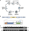
FIG 1
FIG 1
Upregulation of bd3285 during prey invasion by Bdellovibrio bacteriovorus and schematic of the encoded protein. (A) Predatory life cycle of Bdellovibrio bacteriovorus. (1) Attack-phase (AP) B. bacteriovorus cells swim or glide to locate Gram-negative prey bacteria to which they attach (2) and then physically invade, concurrently rounding the rod-shaped prey cell into a spherical prey “bdelloplast” (3). B. bacteriovorus elongates as a filament within the prey periplasm, consuming the nutrients of the dead prey (4) until nutrients are exhausted and the predator divides to yield multiple progeny cells (5). B. bacteriovorus progeny then lyse the dead prey cell and seek out new prey to invade (6). (B) Genetic locus of the monocistronic bd3285 gene within the genome of B. bacteriovorus HD100 (top) and a schematic of the predicted domains and regions of the Bd3285 protein (bottom). Lipo-SP, lipoprotein signal peptide; 3D domain, domain containing 3 aspartate residues (D275, D309, and D321) predicted to be critical for catalysis based on homology to E. coli K-12 MG1655 MltA. (C) Reverse transcriptase PCR performed on B. bacteriovorus HD100 RNA isolated at time points during a predatory cycle on E. coli S17-1 prey. dnaK is a constitutively transcribed control gene. L, molecular weight 100 bp ladder; AP, attack-phase predators; 0.25 to 4, hours since predation commenced; NT, no template RNase-free water; Ec, E. coli S17-1 RNA, G, B. bacteriovorus HD100 genomic DNA. Data are representative of three biological repeats.
FIG 2
FIG 2
Deletion of bd3285 gives prey bdelloplasts with differing morphologies from E. coli prey. (A) Predation of B. bacteriovorus HD100 wild-type or Δbd3285 upon E. coli S17-1 prey showing the different morphologies of prey bdelloplasts invaded by each strain. (B) Magnified images highlighting the three different bdelloplast shapes for prey invaded by Δbd3285 compared to uniquely spherical prey generated by WT HD100 invasion. The proportion of bdelloplasts represented by each shape is denoted in the top right of each image. B. bacteriovorus predator cells are labeled in red via the fluorescent fusion of cytoplasmic protein Bd0064-mCherry. Scale bars = 2 μm and images are representative of three biological repeats. (C) Proportion of spherical bdelloplasts (classified as spherical by a circularity value of >0.96 A.U.) invaded by wild-type, Δbd3285, Δbd3285 (WT comp), or Δbd3285 (D321A comp) predators. Error bars represent SE of the mean. Ns, nonsignificant; **, P < 0.0018; ***, P < 0.0004; (one-way ANOVA with Tukey’s multiple-comparison test). (D) Circularity of bdelloplasts invaded by the same strains. Box, 25th to 75th percentiles; line, median; whiskers, Tukey; ns, nonsignificant; ****, P < 0.0001; (Kruskal-Wallis test with Dunn’s multiple-comparison test). For C and D, n = 234 to 670 cells were analyzed at the 1 h predation time point from three biological repeats.
FIG 3
FIG 3
Prey shape transformation by wild-type and Δbd3285 B. bacteriovorus strains. Time-lapse microscopy stills of prey invasion by either B. bacteriovorus HD100 wild-type (A) or Δbd3285 (B) into E. coli S17-1 prey. Three examples of prey invasion for Δbd3285 are shown: (i) a rod rounding up into a spherical bdelloplast; (ii) a rod shortening in length but remaining rod-shaped; and (iii) a dividing E. coli that shortens in length and becomes a dumbbell bdelloplast. Scale bars = 2 μm and examples are representative of three biological repeats.
FIG 4
FIG 4
Bd3285-mCherry localizes to the septum of dividing E. coli cells. Fluorescence microscopy images of E. coli TOP10 containing the fusion of Bd3285-mCherry within the vector pBAD. Cells were induced with 0.2% arabinose for 20 h and then images were acquired of both nondividing cells (A) and dividing cells with a visible constriction at the midcell (B). Scale bars = 2 μm and images are representative of three biological repeats.
FIG 5
FIG 5
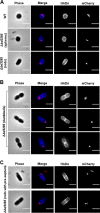
The septum of dividing E. coli is not cleaved in prey invaded by B. bacteriovorus Δbd3285. Fluorescence microscopy images of E. coli S17-1 prey invaded by either B. bacteriovorus wild-type or Δbd3285. The E. coli PG cell wall was prelabeled with the blue D-amino acid HADA prior to predation. B. bacteriovorus predator strains contain a Bd0064-mCherry fusion to label the predator cytoplasm and allow visualization of predators inside prey. Samples were fixed for imaging 30 min after predator-prey mixing. (A) Spherical bdelloplast invaded by a wild-type predator (top row), and spherical and rod-shaped bdelloplasts invaded by Δbd3285 (middle and bottom rows, respectively). (B) Examples of dumbbell-shaped bdelloplasts invaded by Δbd3285 which still contain a septum. (C) Examples of rod-shaped bdelloplasts that appear to contain some preseptal PG. Scale bars = 2 μm and images are representative of three biological repeats.
FIG 6
FIG 6
Model for potential Bd3285 activity during wild-type B. bacteriovorus predation. (A) Model of predation of wild-type B. bacteriovorus upon E. coli prey that is either (i) non-dividing or (ii) dividing at the moment of predator invasion. Non-dividing prey does not contain any septal peptidoglycan; therefore, de-cross-linking activity of DacB enzymes alone is sufficient to convert rod-shaped prey into spherical bdelloplasts (i). In dividing prey with septal peptidoglycan, however, Bd3285 lytic transglycosylase action is required to cleave the septum, facilitating full conversion of dividing cells into spherical bdelloplasts (ii). (B) Close-up model of Bd3285 action at the septum during predation on dividing prey cells.

Comment in

  • J Bacteriol. 205.

Similar articles

Cited by

References

    1. Vollmer W, Blanot D, de Pedro MA. 2008. Peptidoglycan structure and architecture. FEMS Microbiol Rev 32:149–167. doi:10.1111/j.1574-6976.2007.00094.x. - DOI - PubMed
    1. Vollmer W, Joris B, Charlier P, Foster S. 2008. Bacterial peptidoglycan (murein) hydrolases. FEMS Microbiol Rev 32:259–286. doi:10.1111/j.1574-6976.2007.00099.x. - DOI - PubMed
    1. Schaub RE, Chan YA, Lee M, Hesek D, Mobashery S, Dillard JP. 2016. Lytic transglycosylases LtgA and LtgD perform distinct roles in remodeling, recycling and releasing peptidoglycan in Neisseria gonorrhoeae. Mol Microbiol 102:865–881. doi:10.1111/mmi.13496. - DOI - PMC - PubMed
    1. Morlot C, Uehara T, Marquis KA, Bernhardt TG, Rudner DZ. 2010. A highly coordinated cell wall degradation machine governs spore morphogenesis in Bacillus subtilis. Genes Dev 24:411–422. doi:10.1101/gad.1878110. - DOI - PMC - PubMed
    1. Jorgenson MA, Chen Y, Yahashiri A, Popham DL, Weiss DS. 2014. The bacterial septal ring protein RlpA is a lytic transglycosylase that contributes to rod shape and daughter cell separation in Pseudomonas aeruginosa. Mol Microbiol 93:113–128. doi:10.1111/mmi.12643. - DOI - PMC - PubMed

Publication types

LinkOut - more resources