. 2023 May;228(5S):S1158-S1178.
doi: 10.1016/j.ajog.2022.11.1283. Epub 2023 Apr 1.

Meconium-stained amniotic fluid

Affiliations

Meconium-stained amniotic fluid

Dahiana M Gallo et al. Am J Obstet Gynecol. 2023 May.

Abstract

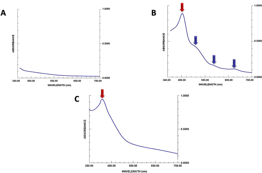
Green-stained amniotic fluid, often referred to as meconium-stained amniotic fluid, is present in 5% to 20% of patients in labor and is considered an obstetric hazard. The condition has been attributed to the passage of fetal colonic content (meconium), intraamniotic bleeding with the presence of heme catabolic products, or both. The frequency of green-stained amniotic fluid increases as a function of gestational age, reaching approximately 27% in post-term gestation. Green-stained amniotic fluid during labor has been associated with fetal acidemia (umbilical artery pH <7.00), neonatal respiratory distress, and seizures as well as cerebral palsy. Hypoxia is widely considered a mechanism responsible for fetal defecation and meconium-stained amniotic fluid; however, most fetuses with meconium-stained amniotic fluid do not have fetal acidemia. Intraamniotic infection/inflammation has emerged as an important factor in meconium-stained amniotic fluid in term and preterm gestations, as patients with these conditions have a higher rate of clinical chorioamnionitis and neonatal sepsis. The precise mechanisms linking intraamniotic inflammation to green-stained amniotic fluid have not been determined, but the effects of oxidative stress in heme catabolism have been implicated. Two randomized clinical trials suggest that antibiotic administration decreases the rate of clinical chorioamnionitis in patients with meconium-stained amniotic fluid. A serious complication of meconium-stained amniotic fluid is meconium aspiration syndrome. This condition develops in 5% of cases presenting with meconium-stained amniotic fluid and is a severe complication typical of term newborns. Meconium aspiration syndrome is attributed to the mechanical and chemical effects of aspirated meconium coupled with local and systemic fetal inflammation. Routine naso/oropharyngeal suctioning and tracheal intubation in cases of meconium-stained amniotic fluid have not been shown to be beneficial and are no longer recommended in obstetrical practice. A systematic review of randomized controlled trials suggested that amnioinfusion may decrease the rate of meconium aspiration syndrome. Histologic examination of the fetal membranes for meconium has been invoked in medical legal litigation to time the occurrence of fetal injury. However, inferences have been largely based on the results of in vitro experiments, and extrapolation of such findings to the clinical setting warrants caution. Fetal defecation throughout gestation appears to be a physiologic phenomenon based on ultrasound as well as in observations in animals.

Keywords: Soret band; bilirubin, biliverdin; discolored amniotic fluid; fetal colonic content, fetal defecation; green-stained amniotic fluid; hypoxia, intraamniotic infection; intraamniotic inflammation; meconium aspiration syndrome; placenta histology; seizures.


Conflict of interest statement

Disclosure: The authors report no conflicts of interest.

Figures

Figure 1.
Figure 1.
Meconium-stained neonate at 39+4 weeks gestation. Figure 1A shows yellow-greenish discoloration of fetal skin at different body sites. The evidence of peripheral cyanosis is shown in Figure 1B (lips), 1C (ears) and 1D (fingertips). Meconium is also present in the ear canal (Figure 1C).
Figure 2.
Figure 2.
The frequency of meconium stained amniotic fluid as a function of gestational age. Modified from Balchin et al
Figure 3.
Figure 3.
Meconium in post maturity syndrome with fetal death at 40 weeks of gestation. The neonate shows the classical features of post maturity syndrome characterized by loss of vernix caseosa, loss of subcutaneous fat and presence of macerated, wrinkled skin (Figures 3A–E). Meconium passage is documented by the greenish-yellow discoloration of the anus, (Figure 3F) the discoloration of the skin (Figure 3A) and the green-yellow staining of placental membranes (Figure 3G).
Figure 4.
Figure 4.
Meconium-stained amniotic fluid. Figure 4A and 4B shows green meconium. Figure 4C and 4D “thin” meconium which is yellow. The traditional concept is that meconium is green when first passed and overtime can become yellow.
Figure 5.
Figure 5.
Spectrophotometric analysis of amniotic fluid. The absorption spectra of amniotic fluid (after centrifugation) with different concentrations of meconium is shown. The band height is linearly correlated with meconium concentration. Modified from Molcho et al
Figure 6.
Figure 6.
The catabolism of heme. Heme is first transformed into biliverdin and then to bilirubin in the reticuloendothelial system. The first reaction consists in the conversion of ferric heme to biliverdin, and it is catalyzed by the heme oxygenase system. Subsequently, biliverdin reductase reduces biliverdin to bilirubin. COOH: carboxyl group; NADPH: Nicotinamide Adenine Dinucleotide Phosphate Hydrogen; NADP+: Nicotinamide adenine dinucleotide phosphate. Modified from Rodwell et al
Figure 7.
Figure 7.
Spectrophotometric analysis of amniotic fluid at different gestational ages. 7A. Normal term amniotic fluid spectrum. The typical spectrum shows a smooth declining slope without peaks suggesting the lack of chemical compounds absorbing light. 7B. Typical spectrophotometric tracing of discolored second-trimester amniotic fluid with a maximum peak near 405nm (“Soret band”, red arrow) and several secondary absorption peaks at 450, 550 and 620 (blue arrows). 7C. Spectrophotometric tracing of term meconium-stained amniotic fluid. There is a peak at 405nm (red arrow) and a smooth declining slope without additional peaks. Modified from Alger et al
Figure 8.
Figure 8.
Chest x-ray showing bilateral patchy opacifications.
Figure 9.
Figure 9.
Autopsy of a neonate with evidence of meconium in the trachea from meconium aspiration syndrome (MAS). The arrow points to fetal hair within the trachea.
Figure 10.
Figure 10.
Meconium in the fetal bronchiole (Figure 9A) and alveoli (Figure 9B). The arrows indicate fetal anucleated squamous cells, one of the components of meconium. Staining is H&E
Figure 11.
Figure 11.
The chorioamniotic membranes in a case of meconium-stained amniotic fluid (stained with H&E). Meconium is visualized within macrophages (blue arrows) in the amnion and chorion stroma (Blue squared parenthesis). Meconium-laden macrophages are recognized by the pink staining of the cytoplasm after excluding hemosiderin pigment with Prussian blue staining (not shown).
Figure 12.
Figure 12.
Chorioamniotic membranes stained with H&E in a case of meconium stained amniotic fluid. Reactive amnion hyperplasia (red arrows) and cytoplasmic vacuolation (blue arrows) are observed.
Figure 13.
Figure 13.
Meconium-induced umbilical cord vessel myonecrosis. 12A. Gross image of the umbilical cord in a case of fetal death with meconium stained amniotic fluid at term. Several areas of ulceration are observed. The Wharton’s jelly is eroded and the vessels are exposed. The dark color represents the muscularis of the vessels (12A). 12B. Shallower ulcerations of the cord. The muscularis is not eroded in this part of the cord. 12C. H&E staining of the umbilical cord. The umbilical vein is on the top and the two arteries below. At this magnification myonecrosis is not evident. 12D. The wall of the umbilical vein with evidence of myonecrosis. 12E. Umbilical artery with damaged myocytes (blue arrows) are observed in the muscular layer closer to the amniotic cavity. Cytoplasmic hypereosinophilia with nuclear pyknosis is evident. The red line indicates the outer perimeter of the umbilical vessel closer to the amniotic cavity, the red arrow indicates the umbilical cord artery lumen. 12F. Cytoplasmic and nuclear changes are better seen at higher magnification (blue arrows in Figure 12F).
Figure 14.
Figure 14.
Evidence of in utero fetal defecation in goats by serial radiographic examinations. After intragastric injection via nasogastric tube, the non-hydrosoluble contrast medium persists in the stomach (red arrow) 4 hours after injection (Figure 13A). Evidence of contrast media in the small bowel (red arrow) 8 hours from injection is shown in figure 13B and eventually the contrast material is excreted in the amniotic cavity (red arrow) where it delineates the fetal body surface and fills the fetal airways. (Figure 13C). Reproduced with permission from Kizilcan et al
Figure 15.
Figure 15.
Results from an experimental study in rabbits performed to investigate the excretion of a radioactive substance (technetium 99, 99mTc-HIDA) injected into the fetal gluteus. The analysis of radioactivity of tissues from fetuses harvested at the rate of one per hour demonstrates that there is physiologic transit of radioactive meconium through the gastrointestinal tract (proximal bowel, mid bowel and distal bowel) into the amniotic fluid. The colored lines represent the magnitude of radioactivity in different tissues. Modified from Ciftci et al
Figure 16.
Figure 16.
Evidence of fetal defecation with 4-Dimensional ultrasound. The anus was examined for 10–15 minutes. Courtesy of López Ramón y Cajal et al
Figure 17.
Figure 17.
Electron microscopic image of fetal cells found in the material from fetal defecation retrieved at amniocentesis. Structures are present on the cell surface that resemble primitive villi (boxed area). Reproduced with permission from López Ramón Y Cajal
Figure 18.
Figure 18.
Meconium debris in the amniotic cavity of twin B (recipient) during fetoscopy in a case of stage III twin to twin transfusion syndrome at 17 weeks. Courtesy of Dr Ramen Chmait.

References

    1. ROSS MG. Meconium aspiration syndrome--more than intrapartum meconium. The New England journal of medicine 2005;353:946–8. - PubMed
    1. GRAND RJ, WATKINS JB, TORTI FM. Development of the human gastrointestinal tract. A review. Gastroenterology 1976;70:790–810. - PubMed
    1. AHANYA SN, LAKSHMANAN J, MORGAN BL, ROSS MG. Meconium passage in utero: mechanisms, consequences, and management. Obstetrical & gynecological survey 2005;60:45–56; quiz 73–4. - PubMed
    1. ROSS WDJA The Works of Aristotle: Historia animalium, by Thompson DW. 1910. Clarendon Press;4;185.
    1. BACK P WALTER K Developmental pattern of bile acid metabolism as revealed by bile acid analysis of meconium. Gastroenterology 1980;78:671–6. - PubMed

Publication types