. 2023 Apr 3;14(1):1863.
doi: 10.1038/s41467-023-37502-z.

Pulmonary inflammation promoted by type-2 dendritic cells is a feature of human and murine schistosomiasis

Affiliations

Pulmonary inflammation promoted by type-2 dendritic cells is a feature of human and murine schistosomiasis

E L Houlder et al. Nat Commun. .

Abstract

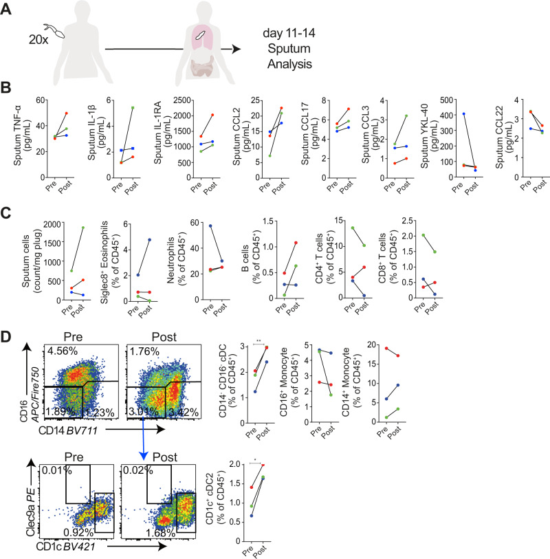
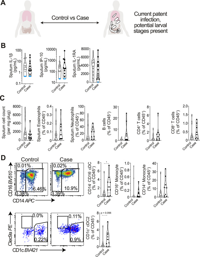
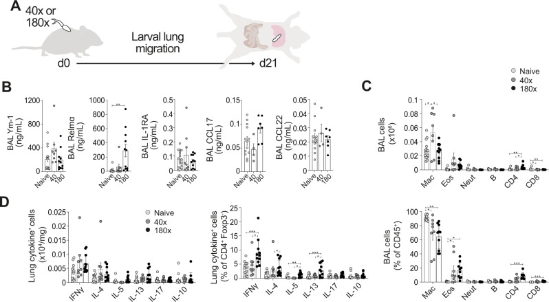
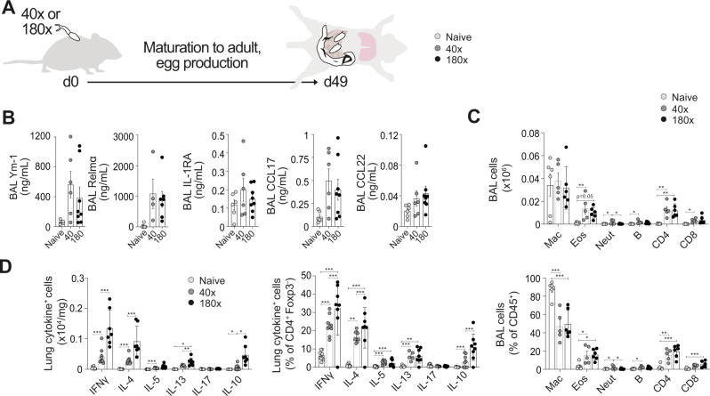
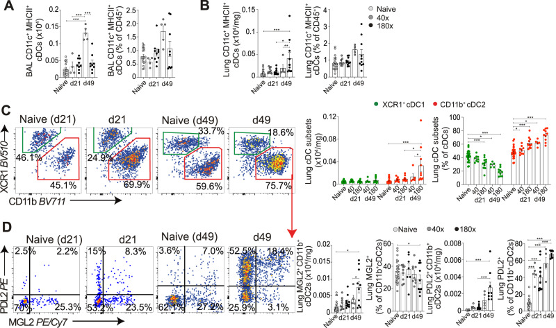
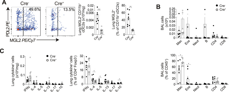
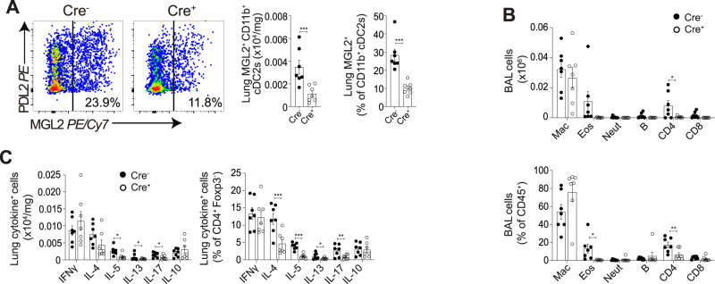
Schistosomiasis is a parasitic disease affecting over 200 million people in multiple organs, including the lungs. Despite this, there is little understanding of pulmonary immune responses during schistosomiasis. Here, we show type-2 dominated lung immune responses in both patent (egg producing) and pre-patent (larval lung migration) murine Schistosoma mansoni (S. mansoni) infection. Human pre-patent S. mansoni infection pulmonary (sputum) samples revealed a mixed type-1/type-2 inflammatory cytokine profile, whilst a case-control study showed no significant pulmonary cytokine changes in endemic patent infection. However, schistosomiasis induced expansion of pulmonary type-2 conventional dendritic cells (cDC2s) in human and murine hosts, at both infection stages. Further, cDC2s were required for type-2 pulmonary inflammation in murine pre-patent or patent infection. These data elevate our fundamental understanding of pulmonary immune responses during schistosomiasis, which may be important for future vaccine design, as well as for understanding links between schistosomiasis and other lung diseases.


Conflict of interest statement

The Manchester Collaborative Centre for Inflammation Research is a joint venture between the University of Manchester and GSK. The authors working at the MCCIR (A.S.M., E.L.H., A.H.C., S.L.B., A.J.L.R., P.C.C., A.A.G., S.A.C.) declare that the research was conducted in the absence of any commercial or financial relationships that could be construed as a potential conflict of interest. The remaining authors declare no competing interests.

Figures

Fig. 1
Fig. 1. Mixed inflammatory responses, and expansion of sputum cDC2s during pre-patent (lung migratory) schistosome infection of non-endemic participants.
A Pulmonary responses were studied in non-endemic participants in Leiden, The Netherlands infected percutaneously with 20 S. mansoni cercariae, with induced sputum samples taken pre-infection, and 11–14 days post-infection. B Inflammatory mediators in sputum supernatants were assessed by Luminex and ELISA. C Sputum cells were isolated, and assessed by flow cytometry for eosinophils, neutrophils, B cells, CD4+ and CD8+ T cells. D Representative flow cytometry plots show changes in monocytes (HLA-DR+, CD14+ or CD16+), cDCs (HLA-DR+, CD14- CD16-), and cDC subsets (CD1c+) following infection. Gate frequencies show % of CD45+ cells. Data are from one study (n = 3 individuals), two-sided paired t tests were used to compare differences between groups. Colours denote individual participants. *P < 0.05, **p < 0.01. Data are presented as mean values ± SEM. Source data are provided as a Source Data file.
Fig. 2
Fig. 2. Muted inflammatory responses and expansion of sputum cDCs in patent schistosome infection in endemic participants.
A Pulmonary responses in uninfected control, and S. mansoni infected cases from Entebbe, Uganda were compared. B Inflammatory mediators in sputum supernatants were assessed by Luminex. Samples below the detection limit were assigned the value of the lowest standard and are shown in blue. Data are from one study (n = 27 individuals). C Sputum cells were isolated, and assessed by flow cytometry for eosinophils, neutrophils, B cells, CD4+ and CD8+ T cells. D Representative flow cytometry plots show alterations in monocytes (HLA-DR+, CD14+ or CD16+), cDCs (HLA-DR+, CD14 CD16-), and cDC subsets (cD1c+) in infected individuals. Gate frequencies show % of CD45+ cells. Data are from one study (n = 21 individuals). Two-sided Mann–Whitney tests were used to compare differences between groups. *p < 0.05. Data are presented as mean values ± SEM. Source data are provided as a Source Data file.
Fig. 3
Fig. 3. Mixed Th2/Th1 pulmonary responses in pre-patent (lung migratory) murine schistosome infection.
A C57BL/6 mice were percutaneously infected with 40 or 180 cercariae, and samples taken at d21. B Inflammatory mediators in BAL were assessed via ELISA. C BAL cell isolates were assessed via flow cytometry for macrophages, eosinophils, neutrophils, B cells, CD4+ and CD8+ T cells. D Lung cell isolates were stimulated with PMA/ionomycin, and cytokine production assessed via flow cytometry. Data are from 4 independent experiments (n = 33 biologically independent animals). Data were fit to a linear mixed effect model, with experimental day as a random effect variable, and groups with a two-sided Tukey’s multiple comparison test. *p < 0.05, **p < 0.01, ***p < 0.001. Data are presented as mean values ± SEM. Source data are provided as a Source Data file.
Fig. 4
Fig. 4. Increased Th2/Th1 pulmonary responses in patent murine schistosome infection.
A C57BL/6 mice were percutaneously infected with 40 or 180 cercariae, and pulmonary samples taken at d49. B Inflammatory mediators in BAL were assessed via ELISA. C BAL cell isolates were assessed via flow cytometry for macrophages, eosinophils, neutrophils, B cells, CD4+ and CD8+ T cells. D Lung cell isolates were stimulated with PMA/ionomycin, and cytokine production assessed via flow cytometry. Data are from 3 independent experiments (n = 26 biologically independent animals). Data were fit to a linear mixed effect model, with experimental day as a random effect variable, and groups compared with a two-sided Tukey’s multiple comparison test. *p < 0.05, **p < 0.01, ***p < 0.001. Data are presented as mean values ± SEM. Source data are provided as a Source Data file.
Fig. 5
Fig. 5. Expansion of pulmonary cDC2s in pre-patent and patent murine schistosome infection.
C57BL/6 mice were percutaneously infected with 40 or 180 cercariae, and samples taken at d21 and d49. Total cDCs (CD11c+MHCII+) were assessed in (A) BAL and (B) lung cell isolates. C Representative flow cytometry plots show cDC subsets (XCR1+ cDC1 and CD11b+ cDC2). Gate frequencies show % of MHCII+ CD11c+ cDCs. D Representative flow cytometry plots show changes in cDC2s. Gate frequencies show % of CD11b+ cDC2s. Double-positive (MGL2+PDL2+) cells are counted as both MGL2+ and PDL2+ in respective graphs. Data are from 7 independent experiments (n = 61 biologically independent animals). Data were fit to a linear mixed effect model, with experimental day as a random effect variable, and groups compared with a two-sided Tukey’s multiple comparison test. *p < 0.05, **p < 0.01, ***p < 0.001. Data are presented as mean values ± SEM. Source data are provided as a Source Data file.
Fig. 6
Fig. 6. Pulmonary IRF4-dependent cDC2s are required to promote pulmonary type-2 immune responses in pre-patent murine schistosome infection.
CD11cΔIrf4 mice were percutaneously infected with 180 cercariae, and samples taken at d21. A Representative flow cytometry plots show depletion of lung MGL2+ IRF4 dependent cDC2s. Gate frequencies show % of CD11b+ cDC2s. B BAL cell isolates were assessed via flow cytometry for macrophages, eosinophils, neutrophils, B cells, CD4+ and CD8+ T cells. C Lung cell isolates were stimulated with PMA/ionomycin, and cytokine production assessed via flow cytometry. Data are from 3 independent experiments (n = 18 biologically independent animals). Data were fit to a linear mixed effect model, with experimental day as a random effect variable, and groups compared with a two-sided LS means Student’s t test. *p < 0.05, **p < 0.01, ***p < 0.001. Data are presented as mean values ± SEM. Source data are provided as a Source Data file.
Fig. 7
Fig. 7. Pulmonary IRF4-dependent cDC2s are required to promote pulmonary type-2 immune responses in patent murine schistosome infection.
CD11cΔIrf4 mice were percutaneously infected with 180 cercariae, and pulmonary samples taken at d49. A Representative flow cytometry plots show depletion of MGL2+ IRF4 dependent cDC2s. Gate frequencies show % of CD11b+ cDC2s. B BAL cell isolates were assessed via flow cytometry for macrophages, eosinophils, neutrophils, B cells, CD4+ and CD8+ T cells. C Lung cell isolates were stimulated with PMA/ionomycin, and cytokine production assessed via flow cytometry. Data are from 2 experiments (n = 14 biologically independent animals). Data were fit to a linear mixed effect model, with experimental day as a random effect variable, and groups compared with a two-sided LS means Student’s t test. *p < 0.05, **p < 0.01, ***p < 0.001. Data are presented as mean values ± SEM. Source data are provided as a Source Data file.

References

    1. Verjee MA. Schistosomiasis: still a cause of significant morbidity and mortality. Res. Rep. Tropical Med. 2020;10:153–163. - PMC - PubMed
    1. Lapa M, et al. Cardiopulmonary manifestations of hepatosplenic schistosomiasis. Circulation. 2009;119:1518–1523. doi: 10.1161/CIRCULATIONAHA.108.803221. - DOI - PubMed
    1. Porres-Aguilar M, Altamirano JT, Torre-Delgadillo A, Charlton MR, Duarte-Rojo A. Portopulmonary hypertension and hepatopulmonary syndrome: a clinician-oriented overview. Eur. Respir. Rev. 2012;21:223–233. doi: 10.1183/09059180.00007211. - DOI - PMC - PubMed
    1. Schwartz E. Pulmonary schistosomiasis. Clin. Chest Med. 2002;23:433–443. doi: 10.1016/S0272-5231(01)00013-2. - DOI - PubMed
    1. Nkurunungi G, et al. Schistosoma mansoni -specific immune responses and allergy in Uganda. Parasite Immunol. 2018;40:e12506. doi: 10.1111/pim.12506. - DOI - PMC - PubMed

Publication types