. 2023 Apr 3;13(1):5425.
doi: 10.1038/s41598-023-32045-1.

Dynamics of inflammatory cytokine expression in bovine endometrial cells exposed to cow blood plasma small extracellular vesicles (sEV) may reflect high fertility

Affiliations

Dynamics of inflammatory cytokine expression in bovine endometrial cells exposed to cow blood plasma small extracellular vesicles (sEV) may reflect high fertility

Pevindu Abeysinghe et al. Sci Rep. .

Abstract

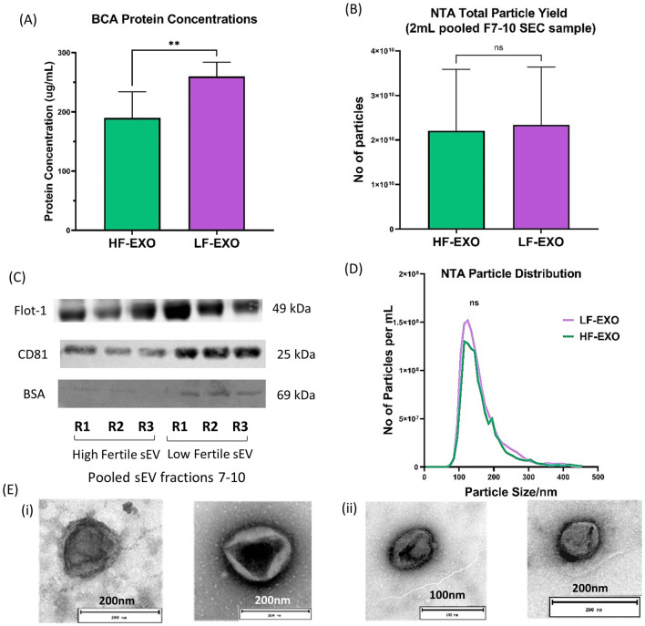
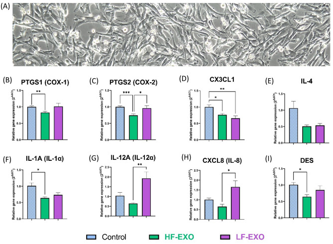
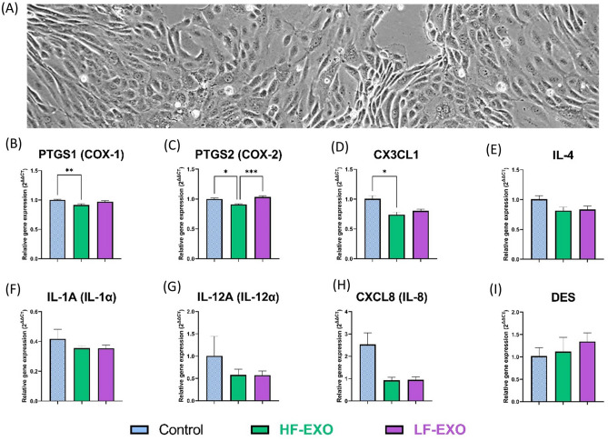
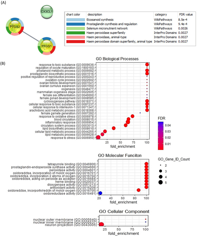
Aberrant inflammation in the endometrium impairs reproduction and leads to poor fertility. Small extracellular vesicles (sEV) are nanoparticles 30-200 nm in-size and contain transferable bioactive molecules that reflect the parent cell. Holstein-Friesian dairy cows with divergent genetic merit, high- (n = 10) and low-fertile (n = 10), were identified based on fertility breeding value (FBV), cow ovulation synchronization and postpartum anovulatory intervals (PPAI). In this study, we evaluated the effects of sEVs enriched from plasma of high-fertile (HF-EXO) and low-fertile (LF-EXO) dairy cows on inflammatory mediator expression by bovine endometrial epithelial (bEEL) and stromal (bCSC) cells. Exposure to HF-EXO in bCSC and bEEL cells yielded lower expression of PTGS1 and PTGS2 compared to the control. In bCSC cells exposed to HF-EXO, pro-inflammatory cytokine IL1-α was downregulated compared to the untreated control, IL-12α and IL-8 were downregulated compared to the LF-EXO treatment. Our findings demonstrate that sEVs interact with both endometrial epithelial and stromal cells to initiate differential gene expression, specifically genes relate to inflammation. Therefore, even subtle changes on the inflammatory gene cascade in the endometrium via sEV may affect reproductive performance and/or outcomes. Further, sEV from high-fertile animals acts in a unique direction to deactivate prostaglandin synthases in both bCSC and bEEL cells and deactivate pro-inflammatory cytokines in the endometrial stroma. The results suggest that circulating sEV may serve as a potential biomarker of fertility.


Conflict of interest statement

The authors declare no competing interests.

Figures

Figure 1
Figure 1
Blood plasma sEV from high fertile and low-fertile both confirmed sEV characteristics and differ in total protein concentrations. (A) Total protein content of sEV from low fertile dairy cow plasma are significantly higher than the high fertile. Values are presented as mean ± SD (**P < 0.001; Mann–Whitney test). (B) However, there’s no significant difference between the particle number (yield) according to the nano-particle tracking analysis (NTA) results. Values are presented as mean ± SD (**P < 0.001; Mann–Whitney test). (C) Representation of western blot sEV/exosome marker CD81, FLOT-1 and negative sEV/exosome marker BSA for pooled sEV fractions 7–10 of HF-EXO and LF-EX. Presence of FLOT-1 and CD81 and less abundance of BSA confirmed sEV/exosome. The full western blot images are available in the Supplementary File 3 (D) The size of sEV (nm) is within the defined size (30–200 nm)—average nano-particle tracking analysis (NTA) particle size distribution of HF-EXO and LF-Exo biological replicates (n = 10 each). There was no statistically significant difference between HF-EXO and LF-EXO particle size distributions (ns represents P > 0.05; Mann–Whitney test). (E) Spherical shape was confirmed in electron micrographs of exosomes from both (i) HF-EXO and (ii) LF-EXO.
Figure 2
Figure 2
Co-incubation with sEV from dairy cows identified as high fertile (HF-EXO) and low fertile (LF-EXO) lead to differential gene expression in bovine endometrial stromal (bCSC) cells. (A) bCSC cells depicts fibroblast-like morphology and grow in multiple layers after confluency (at 48-h, 10X). (B,C) Prostaglandin synthase enzyme gene expression, (D,E) anti-inflammatory mediator gene expression (F–I) pro-inflammatory mediator gene expression in bCSC cells. GAPDH, ACTB and TBP were selected as the house-keeping genes to normalize the qRT-PCR data. Values are presented as mean ± SEM. Brown-Frosythe and Welch ANOVA test along with Dunnett’s T3 multiple comparison was selected as post-hoc test to identify statistical significance differences between the groups. *P ≤ 0.05, **P ≤ 0.01, ***P ≤ 0.001.
Figure 3
Figure 3
Co-incubation with sEV from dairy cows identified as high fertile (HF-EXO) and low fertile (LF-EXO) lead to differential gene expression in bovine endometrial epithelial (bEEL) cells. (A) bEEL cells depicts columnal or cuboidal morphology and showed contact inhibition after confluency (at 48-h, 10X). (B,C) Prostaglandin synthase enzyme gene expression, (D,E) anti-inflammatory mediator gene expression (F,I) pro-inflammatory mediator gene expression in bEEL cells. Values are presented as mean ± SEM. Brown-Frosythe and Welch ANOVA test along with Dunnett’s T3 multiple comparison was selected as post-hoc test to identify statistical significance differences between the groups. *P ≤ 0.05, **P ≤ 0.01, ***P ≤ 0.001.
Figure 4
Figure 4
Gene enrichment analysis for 3 significantly downregulated genes (PTGS1, PTGS2 and CX3CL1) in bovine endometrial cells after incubation with HF-EXO compared to the control (A) STRING protein n-protein interaction (PPI) analysis (B) Gene ontology (GO) enrichment for biological processes, molecular function and cellular component revealed highest enriched GO terms relate to reproduction and inflammation. Fisher’s exact test was used for GO annotations. The corrected P-value was calculated to false discovery rate (FDR), FDR < 0.0001.

References

    1. Mor G, Cardenas I. The immune system in pregnancy: A unique complexity. Am. J. Reprod. Immunol. 2010;63(6):425–33. doi: 10.1111/j.1600-0897.2010.00836.x. - DOI - PMC - PubMed
    1. Naim N, Amrit FRG, McClendon TB, Yanowitz JL, Ghazi A. The molecular tug of war between immunity and fertility: Emergence of conserved signaling pathways and regulatory mechanisms. Bioessays. 2020;42(12):e2000103. doi: 10.1002/bies.202000103. - DOI - PMC - PubMed
    1. Shynlova O, Lee Y-H, Srikhajon K, Lye SJ. Physiologic uterine inflammation and labor onset: Integration of endocrine and mechanical signals. Reprod. Sci. 2013;20(2):154–167. doi: 10.1177/1933719112446084. - DOI - PubMed
    1. Marzi M, Vigano A, Trabattoni D, Villa ML, Salvaggio A, Clerici E, et al. Characterization of type 1 and type 2 cytokine production profile in physiologic and pathologic human pregnancy. Clin. Exp. Immunol. 1996;106(1):127–33. doi: 10.1046/j.1365-2249.1996.d01-809.x. - DOI - PMC - PubMed
    1. Chatterjee P, Chiasson VL, Bounds KR, Mitchell BM. Regulation of the anti-inflammatory cytokines interleukin-4 and interleukin-10 during pregnancy. Front. Immunol. 2014 doi: 10.3389/fimmu.2014.00253. - DOI - PMC - PubMed

Publication types