. 2023 Jul;31(7):784-792.
doi: 10.1038/s41431-023-01320-0. Epub 2023 Apr 4.

The genetic landscape and clinical implication of pediatric Moyamoya angiopathy in an international cohort

Affiliations

The genetic landscape and clinical implication of pediatric Moyamoya angiopathy in an international cohort

Paolo Zanoni et al. Eur J Hum Genet. 2023 Jul.

Abstract

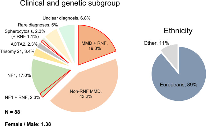
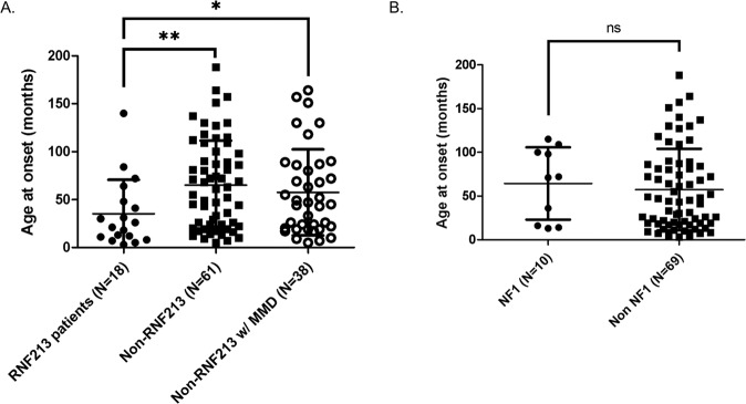
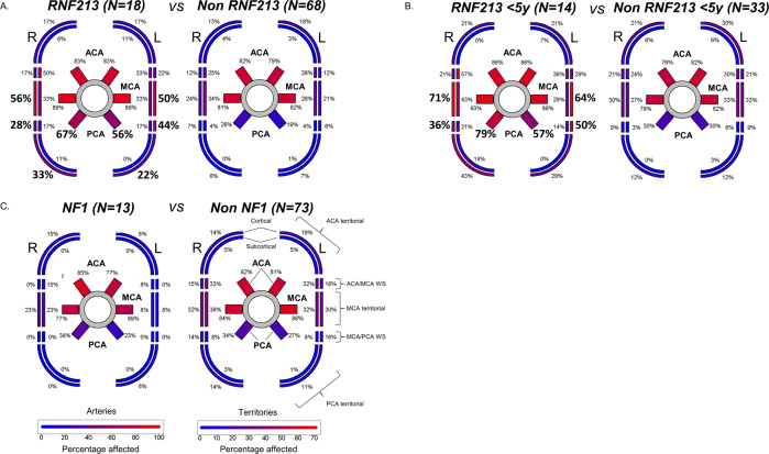
Pediatric Moyamoya Angiopathy (MMA) is a progressive intracranial occlusive arteriopathy that represents a leading cause of transient ischemic attacks and strokes in childhood. Despite this, up to now no large, exclusively pediatric MMA cohort has been subjected to systematic genetic investigation. In this study, we performed molecular karyotyping, exome sequencing and automated structural assessment of missense variants on a series of 88 pediatric MMA patients and correlated genetic, angiographic and clinical (stroke burden) findings. The two largest subgroups in our cohort consisted of RNF213 and neurofibromatosis type 1 (NF1) patients. While deleterious RNF213 variants were associated with a severe MMA clinical course with early symptom onset, frequent posterior cerebral artery involvement and higher stroke rates in multiple territories, NF1 patients had a similar infarct burden compared to non-NF1 individuals and were often diagnosed incidentally during routine MRIs. Additionally, we found that MMA-associated RNF213 variants have lower predicted functional impact compared to those associated with aortic disease. We also raise the question of MMA as a feature of recurrent as well as rare chromosomal imbalances and further support the possible association of MMA with STAT3 deficiency. In conclusion, we provide a comprehensive characterization at the genetic and clinical level of a large exclusively pediatric MMA population. Due to the clinical differences found across genetic subgroups, we propose genetic testing for risk stratification as part of the routine assessment of pediatric MMA patients.


Conflict of interest statement

The authors declare no competing interests.

Figures

Fig. 1
Fig. 1. Clinical presentation and genetic subgroups of the study cohort.
Main clinical and genetic subgroups (left) and ethnicity (right). The percentage of cases with predicted deleterious and possibly deleterious RNF213 variants (as defined in Fig. 2B) are highlighted in red and labeled as “RNF”. In the left panel, “rare diagnoses” were SHAM syndrome, Alagille syndrome, MOPD type 2 syndrome, STAT3 deficiency and brain small vessel disease type 2. “Unclear diagnosis” refers to MMS patients for whom neither a clinical nor a genetic diagnosis associated with MMA could be established.
Fig. 2
Fig. 2. Location and pathogenicity prediction of RNF213 variants.
A Variant location. The diagram shows the structure of the RNF213 gene together with the 25 variants identified in our study population (in bold; colored for pathogenicity scores as in panel 2B) as well as previously reported variants detected either in patients with MMA /intracranial arterial stenoses (above) or with other arteriopathies (below; source: HGMD (http://www.hgmd.cf.ac.uk/ac/index.php)). Previously reported variants that have been detected in our study population as well are underlined. Amino acid coordinates refer to transcript NM_001256071.3; for simplicity, the one-letter code was used to symbolize amino acids; for HGVS-conform variant nomenclature, refer to Tables S3 and S4; domain locations were derived from Ahel et al. [8]. N-arm: N-terminal arm; RING: RING finger domain; CTD: C-terminal domain. B Variant classification based on the combination of CADD and VIPUR scores. All variants detected in our cohort except G405del, which could not be tested using the VIPUR algorithm, are shown. Density distribution of CADD and VIPUR scores are displayed on the side of each respective axis. Data points were classified by expectation-maximization as described in the methods. Please refer to the legend on top of the graph for color coding of the variants. All heterozygotes for predicted deleterious and possibly deleterious variant are described as “RNF213 patients” in this paper. C Comparison with previously reported RNF213 variants. The VIPUR and CADD scores of 90 previously reported RNF213 variants (source: HGMD) are shown here together with the variants detected in this study (see A; labeled as “MMA – this study”). Data points were colored based on the reported clinical phenotype. For 11 previously reported variants no VIPUR score could be generated either because they were located in the N-terminal region (no structural information available; 8 variants) or they were nonsense (2 variants) or synonymous (1 variant). Each point represents one variant. Segments represent the distance between each variant and the respective group’s average. Ellipses represent data distribution in each group assuming a multivariate normal distribution with a confidence threshold of 95%.
Fig. 3
Fig. 3. Correlation between genotype and age at onset in our cohort.
A Age at MMA symptom onset for RNF213 patients compared to all non-RNF213 as well as to all non-RNF213 with MMD. B Age at MMA symptom onset for patients with a clinical diagnosis of NF1 compared to all other patients in our cohort. Patients diagnosed by MRI in the asymptomatic phase as well as NF1 patients with additional RNF213 (possibly) deleterious variants were excluded from this analysis. Lines and whiskers represent mean ± SD. ns non-significant, *p < 0.05, **p < 0.01 by two-tailed Mann–Whitney test.
Fig. 4
Fig. 4. Genotype–phenotype correlation for angiographic and MRI findings.
Schematic representation of the Circle of Willis and the respective brain territories supplied by the anterior, middle and posterior cerebral artery (ACA, MCA and PCA). Arteries involved and their respective territories are colored based on percentage of patients affected, as reported in the legend at the bottom. The involvement of each artery and territory was compared between RNF213 patients (A), RNF213 patients below the age of 5 (B), NF1 patients (C) and the respective rest of the cohort by Fischer’s Exact Test (FET) irrespective of laterality. Significant differences (FET p value <0.05 after Bonferroni correction) are shown in bold. Patients aged below 5 years (C) were separately analyzed in order to exclude the possible confounding effect of age, as RNF213 patients are younger than non-RNF213 ones (Fig. 3A). Two NF1 patients (68632 and 82059) were excluded from this analysis as they carried an additional (possibly) deleterious RNF213 variant as well.

Comment in

References

    1. Scott RM, Smith ER. Moyamoya disease and moyamoya syndrome. N Engl J Med. 2009;360:1226–37. doi: 10.1056/NEJMra0804622. - DOI - PubMed
    1. Kim SK, Cho BK, Phi JH, Lee JY, Chae JH, Kim KJ, et al. Pediatric moyamoya disease: an analysis of 410 consecutive cases. Ann Neurol. 2010;68:92–101. doi: 10.1002/ana.21981. - DOI - PubMed
    1. Kim JS. Moyamoya disease: epidemiology, clinical features, and diagnosis. J Stroke. 2016;18:2–11. doi: 10.5853/jos.2015.01627. - DOI - PMC - PubMed
    1. Kleinloog, R, Regli, L, Rinkel, GJE, Klijn, CJM. Regional differences in incidence and patient characteristics of moyamoya disease: a systematic review. J Neurol Neurosurg Psychiatry. 2012;83:531–6. http://jnnp.bmj.com/. - PubMed
    1. Kuroda S, Fujimura M, Takahashi J, Kataoka H, Ogasawara K, Iwama T, et al. Diagnostic criteria for moyamoya disease-2021 revised version. Neurol Med Chir. 2022;62:307–12. doi: 10.2176/jns-nmc.2022-0072. - DOI - PMC - PubMed

Publication types

Substances