. 2023 Apr 3;6(1):365.
doi: 10.1038/s42003-023-04661-0.

LncAABR07053481 inhibits bone marrow mesenchymal stem cell apoptosis and promotes repair following steroid-induced avascular necrosis

Affiliations

LncAABR07053481 inhibits bone marrow mesenchymal stem cell apoptosis and promotes repair following steroid-induced avascular necrosis

Tao Wang et al. Commun Biol. .

Abstract

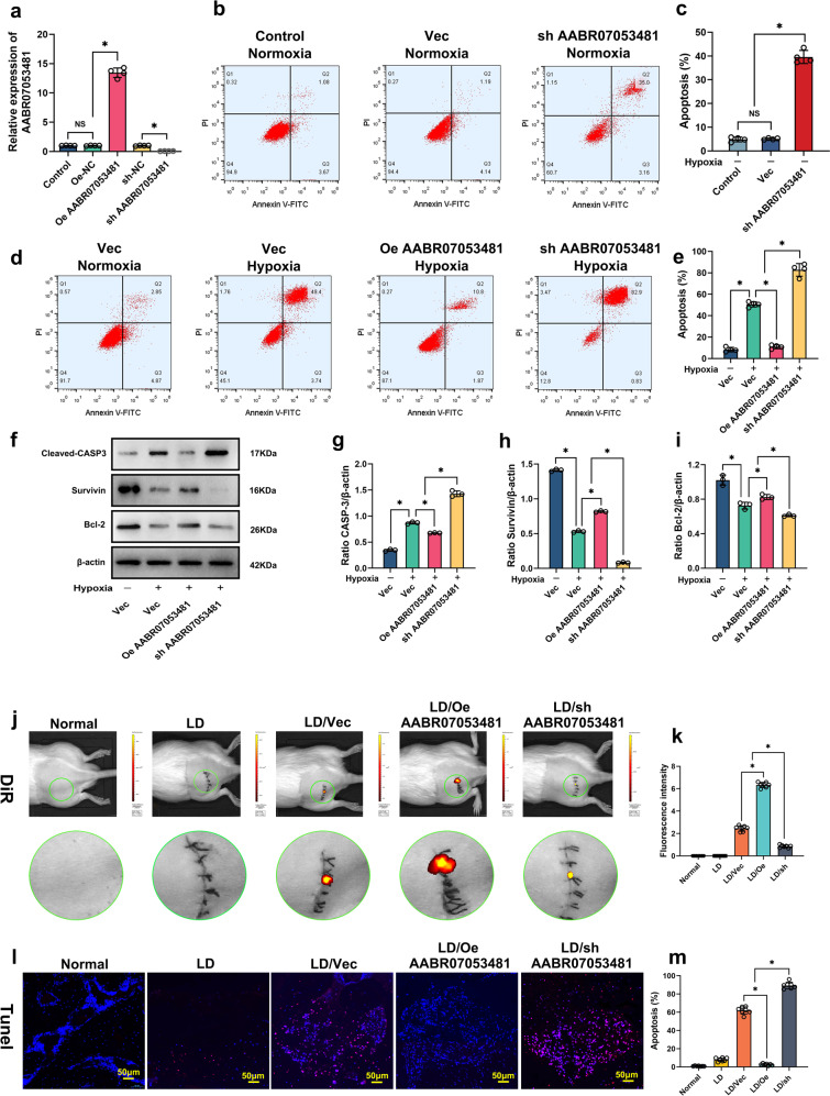
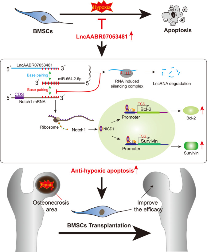
The osteonecrotic area of steroid-induced avascular necrosis of the femoral head (SANFH) is a hypoxic microenvironment that leads to apoptosis of transplanted bone marrow mesenchymal stem cells (BMSCs). However, the underlying mechanism remains unclear. Here, we explore the mechanism of hypoxic-induced apoptosis of BMSCs, and use the mechanism to improve the transplantation efficacy of BMSCs. Our results show that the long non-coding RNA AABR07053481 (LncAABR07053481) is downregulated in BMSCs and closely related to the degree of hypoxia. Overexpression of LncAABR07053481 could increase the survival rate of BMSCs. Further exploration of the downstream target gene indicates that LncAABR07053481 acts as a molecular "sponge" of miR-664-2-5p to relieve the silencing effect of miR-664-2-5p on the target gene Notch1. Importantly, the survival rate of BMSCs overexpressing LncAABR07053481 is significantly improved after transplantation, and the repair effect of BMSCs in the osteonecrotic area is also improved. This study reveal the mechanism by which LncAABR07053481 inhibits hypoxia-induced apoptosis of BMSCs by regulating the miR-664-2-5p/Notch1 pathway and its therapeutic effect on SANFH.


Conflict of interest statement

The authors declare no competing interests.

Figures

Fig. 1
Fig. 1. LncAABR07053481 is downregulated under hypoxia and associated with apoptosis of BMSCs.
a Cluster analysis of LncRNAs (n = 3). b Location of LncRNA in the genome. c Prediction of LncAABR07053481 coding capability by the coding-potential assessment tool (CPAT). d The expression of the Myc-fused protein was analyzed by immunoblotting with an anti-Myc antibody (n = 3). e, f The relationship between the degree of hypoxia and apoptosis (n = 4). g Expression of LncAABR07053481 under different oxygen concentrations (n = 4). h Detection of LncAABR07053481 expression in the normal femoral head and the area of osteonecrosis by qPCR (n = 6). i Detection of LncAABR07053481 expression in BMSCs at different times after transplantation by qPCR (n = 6). Data were shown as mean ± SD. *P < 0.05; In (h), statistical significance was calculated by Student’s t-test; in (f, g, i), statistical significance was calculated by one-way ANOVA with Tukey’s post hoc tests.
Fig. 2
Fig. 2. LncAABR07053481 can inhibit hypoxia-induced apoptosis of BMSCs.
a The expression of LncAABR07053481 was detected by qPCR (n = 4). be Detection of BMSC apoptosis under different conditions by Annexin V-fluorescein isothiocyanate (FITC, green)/propidium iodide (PI, red) (n = 4). fi The expression levels of CASP-3, Survivin, and Bcl-2 were detected by western blotting (n = 3). j Forty-eight hours after surgery, the fluorescence intensity of 1,1-dioctadecyl-3,3,3,3-tetramethylindotricarbocyanine iodide (DiR, red) in the transplantation area was detected by live imaging of small animals (n = 7). k Quantitative analysis of DiR fluorescence intensity in the transplanted area (n = 7). lm At 48 h after surgery, TdT-mediated dUTP nick-end labeling (TUNEL, red) /4’,6-diamidino-2-phenylindole (DAPI, blue) was used to detect apoptosis in the transplantation area (n = 7). Data were shown as mean  ±  S.D. *P < 0.05; in (a, c, e, gi, k, m), statistical significance was calculated by one-way ANOVA with Tukey’s post hoc test.
Fig. 3
Fig. 3. LncAABR07053481 inhibits hypoxia-induced apoptosis of BMSCs by regulating the Notch1 pathway.
a mRNA clustering analysis (n = 3). b Kyoto Encyclopedia of Genes and Genomes (KEGG) pathway enrichment analysis of differentially expressed genes. c Gene set enrichment analysis (GSEA). d The expression of Notch1 was detected by qPCR (n = 3). eg The expression levels of Notch1 and NICD1 were detected by western blotting (n = 3). h, i Detection of BMSC apoptosis by Annexin V-FITC/PI (n = 3). jn The expression levels of Notch1, NICD1, Survivin, and Bcl-2 were detected by western blotting (n = 3). Data were shown as mean  ±  SD. *P < 0.05; In (d, f, g), statistical significance was calculated by Student’s t-test; In (i, kn), statistical significance was calculated by one-way ANOVA with Tukey’s post hoc test.
Fig. 4
Fig. 4. LncAABR07053481 functions as a ceRNA and sponges miR-664-2-5p in BMSCs.
a RNA fluorescence in situ hybridization (RNA-FISH) detection of the subcellular localization of LncAABR07053481, 18S, and U6 were used as positive control. (n = 4). b Bioinformatics tools predict the miRNAs that may bind to the untranslated region at the 3’ end of Notch1 mRNA (Notch1 mRNA 3′UTR) and LncAABR07053481. c Binding sites of miR-664-2-5p with LncAABR07053481 and Notch1 mRNA 3′UTR. d, e Luciferase reporter assay shows that miR-664-2-5p binds to LncAABR07053481 and Notch1 mRNA (n = 5). fh RNA immunoprecipitation (RIP) detection of LncAABR07053481 enrichment to miRNA ribonucleoprotein complexes (miRNPs) (n = 4). gi RIP detection of enrichment of Notch1 mRNA to miRNPs (n = 4). Data were shown as mean  ±  SD. *P < 0.05; In (di), statistical significance was calculated by one-way ANOVA with Tukey’s post hoc test.
Fig. 5
Fig. 5. The LncAABR07053481/miR-664-2-5p/Notch1 axis is involved in the regulation of hypoxia-induced apoptosis of BMSCs.
ac The expressions of LncAABR07053481, miR-664-2-5p, and NICD1 were detected by qPCR (n = 4). df The expression levels of Notch1 and NICD1 were detected by western blotting (n = 3). g, h Detection of BMSC apoptosis under different conditions by TUNEL/DAPI (n = 3). Data were shown as mean  ± SD. *P < 0.05; In (ac, e, f, h), statistical significance was calculated by one-way ANOVA with Tukey’s post hoc test.
Fig. 6
Fig. 6. LncAABR07053481-overexpressed BMSCs promote the repair of early SANFH.
a At 12 weeks after BMSC transplantation, micro-CT was used to analyze the repair of the necrotic area of the femoral head (n = 6). bd Quantitative analysis of the number of trabeculae, the trabecular thickness, and the volume fraction of new bone tissue (n = 6). e At 12 weeks after BMSC transplantation, H&E and Masson staining were used to evaluate the repair of the necrotic area of the femoral head (n = 6). f At 12 weeks after BMSC transplantation, immunohistochemistry was used to detect osteogenic markers of runt-related transcription factor 2 (Runx2, Cy3 label, red) and osteopontin (OPN, FITC label, green) (n = 6). g Quantitative analysis of Runx2 expression (n = 6). h Quantitative analysis of OPN expression (n = 6). Data were shown as mean ± SD. *P < 0.05; In (bd, g, h), statistical significance was calculated by one-way ANOVA with Tukey’s post hoc test.
Fig. 7
Fig. 7. Effect and mechanism of LncAABR07053481 on the repair of early SANFH by regulating the Notch1 pathway to inhibit hypoxia-induced apoptosis of BMSCs: LncAABR07053481 acts as a sponge for miR-664-2-5p to attenuate its repressive effect on Notch1 and activates the Notch1 pathway and releases NICD1 (the intracellular active fragment of Notch1) from the cell membrane.
NICD1 localizes to the nucleus, where it forms a transcriptionally active complex with the DNA-binding protein CSL and the coactivator Mastermind (MAM) to upregulate transcription of Notch target genes (Survivin and Bcl-2). Following which, hypoxia-induced apoptosis of BMSCs in the area of osteonecrosis is inhibited, improving the transplantation effect of BMSCs on early SANFH.

References

    1. Weinstein RS. Glucocorticoid-induced osteonecrosis. Endocrine. 2012;41:183–190. doi: 10.1007/s12020-011-9580-0. - DOI - PMC - PubMed
    1. Xie XH, Wang XL, Yang HL, Zhao DW, Qin L. Steroid-associated osteonecrosis: epidemiology, pathophysiology, animal model, prevention, and potential treatments (an overview) J. Orthop. Transl. 2015;3:58–70. - PMC - PubMed
    1. Chang C, Greenspan A, Gershwin ME. The pathogenesis, diagnosis and clinical manifestations of steroid-induced osteonecrosis. J. Autoimmun. 2020;110:102460. doi: 10.1016/j.jaut.2020.102460. - DOI - PubMed
    1. Krez A, et al. Risk factors for multi-joint disease in patients with glucocorticoid-induced osteonecrosis. Osteoporos. Int. 2021;20:1–9. - PMC - PubMed
    1. Wang A, Ren M, Wang J. The pathogenesis of steroid-induced osteonecrosis of the femoral head: a systematic review of the literature. Gene. 2018;671:103–109. doi: 10.1016/j.gene.2018.05.091. - DOI - PubMed

Publication types