

doi: 10.1016/j.eats.2022.11.023.
eCollection 2023 Mar.
Three-Dimensional Printing of the Patellofemoral Joints of Patellar Instability Patients
Affiliations
- PMID: 37013007
- PMCID: PMC10066413
- DOI: 10.1016/j.eats.2022.11.023
Item in Clipboard
Three-Dimensional Printing of the Patellofemoral Joints of Patellar Instability Patients
Arthrosc Tech.
.
Abstract
Three-dimensional (3D) modeling and printing comprise an important tool for orthopaedic surgeons. One area in which 3D modeling has the potential to dramatically improve our understanding of biomechanical kinematics is pathologies of the patellofemoral joint, in particular trochlear dysplasia. We describe a method for creating 3D printed models of the patellofemoral joint, including computed tomography image acquisition, image segmentation, model creation, and 3D printing. The models created can help surgeons understand and plan surgery for recurrent patellar dislocations.
© 2022 The Authors.
Figures

Weight-bearing computed tomography (CT) scan acquisition. The first step of creating a 3-dimensional (3D) model of the patellofemoral joint is imaging the patient. The Carestream Onsight 3D Extremity system is used to capture a weight-bearing CT scan of the knee. The patient is asked to stand with 1 leg in the machine, after which the door is closed and the height of the machine is adjusted. The patient places his or her arms on the support bar in a neutral standing position for scout localizer image and scan acquisition. After the acquisition of scans in the neutral position, the patient remains standing but flexes the knee to 20° of flexion. Each CT acquisition takes approximately 1 minute.

Three-dimensional (3D) print generation from computed tomography files. (A) To convert imaging data to a 3D model using Simpleware ScanIP, mask thresholding and segmentation are performed by selecting a range of Hounsfield units to be included in the model of the distal femur using the thresholding tool. The range should be optimized to include only bone, without extraneous signal in soft tissue. Suboptimal thresholding is shown, requiring further alteration of the Hounsfield unit range applied. (B) The rendered 3D reconstruction can be cropped to highlight the anatomic region of interest by altering window visibility in each direction (right, left, anterior, posterior, and so on).
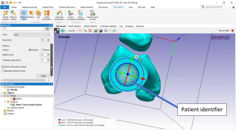
The model of the distal femur is prepared for printing and given a unique embossed label for identification after the printing process. After this preparation in ScanIP, the model is exported as an STL file for printing.

Model importation for printing. The STL file of the 3-dimensional model of the distal femur is imported into PreForm, a software platform compatible with the Formlabs family of 3-dimensional printers, and oriented on the baseplate for printing.

Three-dimensional print curing under ultraviolet light. After completion of the 3-dimensional printing process, model washing in isopropyl alcohol, and a 30-minute drying period, the 3-dimensionally printed distal femur is placed in the Form Cure system for ultraviolet treatment for 30 minutes. After completion of curing, any support struts present are removed and the model is considered ready for analysis.

Completed 3-dimensionally printed model of distal femur, which can now be used to better understand patient’s trochlear morphology.
References
-
- Bagaria V., Chaudhary K. A paradigm shift in surgical planning and simulation using 3Dgraphy: Experience of first 50 surgeries done using 3D-printed biomodels. Injury. 2017;48:2501–2508. - PubMed
-
- Chi-Kay L., King-him C., Kin-bong L., Wilson L. Computer-assisted planning and three-dimensional-printed patient-specific instrumental guide for corrective osteotomy in post-traumatic femur deformity: A case report and literature review. J Orthop Trauma Rehabil. 2018;24:12–17.
-
- Lin C.Y., Wirtz T., LaMarca F., Hollister S.J. Structural and mechanical evaluations of a topology optimized titanium interbody fusion cage fabricated by selective laser melting process. J Biomed Mater Res A. 2007;83:272–279. - PubMed
-
- Koeck F.X., Beckmann J., Luring C., Rath B., Grifka J., Basad E. Evaluation of implant position and knee alignment after patient-specific unicompartmental knee arthroplasty. Knee. 2011;18:294–299. - PubMed
LinkOut - more resources
Full Text Sources
