Case Reports
. 2022 Nov;12(11):87-90.
doi: 10.13107/jocr.2022.v12.i11.3426.

Post-traumatic Osteomyelitis of the Rib-point of Care In children, Presenting with chest Wall Pain

Affiliations
Case Reports

Post-traumatic Osteomyelitis of the Rib-point of Care In children, Presenting with chest Wall Pain

Pankaj Kumar Mishra et al. J Orthop Case Rep. 2022 Nov.

Abstract

Introduction: The rib osteomyelitis is a very rare entity and it hardly accounts for 1% of all cases of osteomyelitis. In this case report, we are presenting a case of acute osteomyelitis of rib in a very young child, with a previous history of mediocre trauma over the chest wall.

Case report: It is a case report of a young boy, who had sustained the blunt injury over the chest wall. The X-ray was unremarkable. After sometime, he presented to the hospital with the pain over the chest wall. Now, the X-ray showed the signs of rib osteomyelitis.

Conclusion: In children, the clinical presentation of rib osteomyelitis is very non-specific. Sometimes, the injury while playing, which is very usual in this age group may create the confusion. Hence, it may need high index of suspicion by the physician to include is as a possible diagnosis.

Keywords: Rib; chest wall; osteomyelitis.


Conflict of interest statement

Conflict of Interest: Nil

Figures

Figure 1
Figure 1
X-ray (posteroanterior view) of the chest of the child, at the first visit after the alleged history of blunt trauma over the left side of the anterior chest wall. X-ray appeared near normal, and he was managed by the presumptive diagnosis of soft-tissue injury.
Figure 2
Figure 2
Now, after 3 month of previous trauma, the X-ray shows that the left 10th rib was swollen, deformed, destructed and full of sequestrum, and suggestive of osteomyelitis.
Figure 3
Figure 3
The magnetic resonance examination revealed that, the lesion was hypointence in T1 (a) and hyperintence in T2 (b) weighted MRI, with thin internal septations and suggestive of rib osteomyelitis.
Figure 4
Figure 4
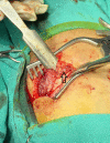
Clinical image, showing the drainage of pus (arrow).
Figure 5
Figure 5
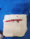
The image showing the excised sequestrum from the rib.
Figure 6
Figure 6
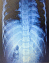
X-ray chest (AP view) showing the healed osteomyelitis of rib, at the end of one year of follow-up.

References

    1. Dich VQ, Nelson JD, Haltalin KC. Osteomyelitis in infants and children. A review of 163 cases. Am J Dis Child. 1975;129:1273–8. - PubMed
    1. Peltola H, Pääkkönen M. Acute osteomyelitis in children. N Engl J Med. 2014;370:352–60. - PubMed
    1. Kashyap NK, Jindal A, Borkar NK, Wasnik M. Primary tuberculotic osteomyelitis of rib in a child. J Clin Diagn Res. 2017;11:D08–9. - PMC - PubMed
    1. Annen MJ, Johnston MJ, Gormley JP, Silverman E. Acute hematogenous osteomyelitis presenting as a “Cold”rib in a child. World J Nucl Med. 2017;16:160–2. - PMC - PubMed
    1. Osinowo O, Adebo OA, Okubanjo AO. Osteomyelitis of the ribs in Ibadan. Thorax. 1986;41:58–60. - PMC - PubMed

Publication types

LinkOut - more resources