. 2023 Apr 3;18(1):35.
doi: 10.1186/s13020-023-00739-3.

Liquiritin exhibits anti-acute lung injury activities through suppressing the JNK/Nur77/c-Jun pathway

Affiliations

Liquiritin exhibits anti-acute lung injury activities through suppressing the JNK/Nur77/c-Jun pathway

Hongling Zhou et al. Chin Med. .

Abstract

Background: Licorice (Glycyrrhiza uralensis Fisch.), a well-known traditional medicine, is traditionally used for the treatment of respiratory disorders, such as cough, sore throat, asthma and bronchitis. We aim to investigate the effects of liquiritin (LQ), the main bioactive compound in licorice against acute lung injury (ALI) and explore the potential mechanism.

Methods: Lipopolysaccharide (LPS) was used to induce inflammation in RAW264.7 cells and zebrafish. Intratracheal instillation of 3 mg/kg of LPS was used for induction an ALI mice model. The concentrations of IL-6 and TNF-α were tested using the enzyme linked immunosorbent assay. Western blot analysis was used to detect the expression of JNK/Nur77/c-Jun related proteins. Protein levels in bronchoalveolar lavage fluid (BALF) was measured by BCA protein assay. The effect of JNK on Nur77 transcriptional activity was determined by luciferase reporter assay, while electrophoretic mobility shift assay was used to examine the c-Jun DNA binding activity.

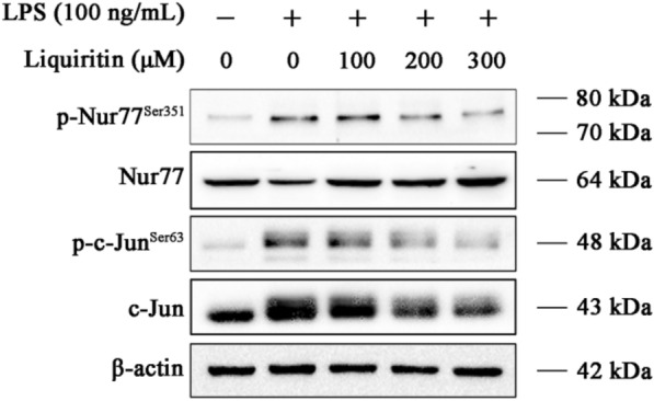
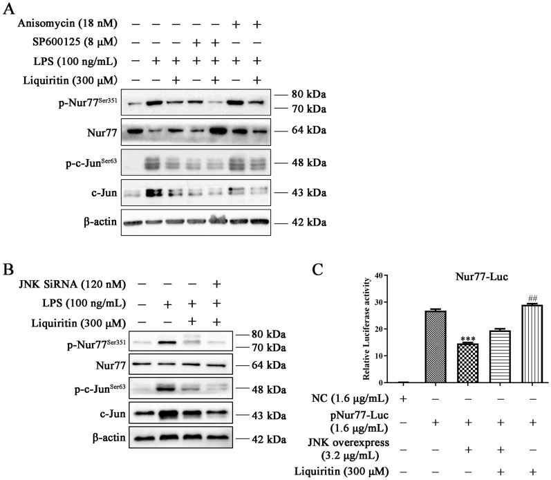
Results: LQ has significant anti-inflammatory effects in zebrafish and RAW264.7 cells. LQ inhibited the expression levels of p-JNK (Thr183/Tyr185), p-Nur77 (Ser351) and p-c-Jun (Ser63), while elevated the Nur77 expression level. Inhibition of JNK by a specific inhibitor or small interfering RNA enhanced the regulatory effect of LQ on Nur77/c-Jun, while JNK agonist abrogated LQ-mediated effects. Moreover, Nur77-luciferase reporter activity was suppressed after JNK overexpression. The effects of LQ on the expression level of c-Jun and the binding activity of c-Jun with DNA were attenuated after Nur77 siRNA treatment. LQ significantly ameliorated LPS-induced ALI with the reduction of lung water content and BALF protein content, the downregulation of TNF-α and IL-6 levels in lung BALF and the suppression of JNK/Nur77/c-Jun signaling, which can be reversed by a specific JNK agonist.

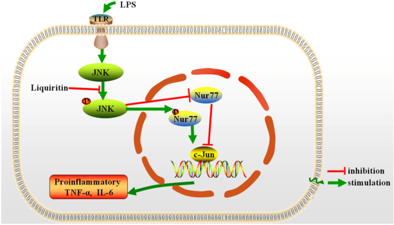
Conclusion: Our results indicated that LQ exerts significant protective effects against LPS-induced inflammation both in vivo and in vitro via suppressing the activation of JNK, and consequently inhibiting the Nur77/c-Jun signaling pathway. Our study suggests that LQ may be a potential therapeutic candidate for ALI and inflammatory disorders.

Keywords: Acute lung injury; JNK; Lipopolysaccharide; Liquiritin; Nur77; c-Jun.


Conflict of interest statement

The authors declare that they have no competing interests.

Figures

Fig. 1
Fig. 1
LQ has significant anti-inflammatory effects in vitro and in vivo. A The chemical structure of LQ. B LQ improved the survival rate of LPS-microinjected zebrafish larvae. One hundred and eighty larvae were randomly divided into 6 groups (n = 30). larvae were exposed to indicated dosages of LQ after PBS or LPS (2 mg/mL) yolks microinjection, then the mortalities of the larvae were observed for 72 h. C Representative fluorescence images and (D) statistical analysis of macrophages in yolk were shown. E RAW264.7 cells viability was determined by MTT assay. F RAW264.7 macrophage cells were pretreated with LQ (100, 200, 300 µM) for 2 h, then LPS (100 ng/mL) were added for another 22 h incubation. The supernatants were collected and the concentrations of TNF-α and IL-6 were determined by ELISA. Data were presented as mean ± SEM, n = 3, ###P < 0.001 versus control; *P < 0.05 versus LPS, **P < 0.01 versus LPS, ***P < 0.001 versus LPS. One-way ANOVA, post hoc comparisons, Turkey’s test
Fig. 2
Fig. 2
LQ exerts anti-inflammatory effects through inhibiting JNK. A LQ inhibited the phosphorylation of JNK. RAW264.7 macrophage cells were treated with LQ (100–300 µM) for 2 h, then LPS (100 ng/mL) were treated for another 22 h. Protein expression levels of p-JNK, JNK, p-ERK, ERK, p-p38 and p38 were determined by Western blotting. B LQ inhibits the pro-inflammatory cytokines by suppressing of JNK. RAW264.7 cells were treated with JNK inhibitor SP600125 (70 nM) or JNK activator anisomycin (18 nM) for 2 h before the administration of LQ, and treated with LPS for another 22 h. Cell supernatants were collected, and the concentration of TNF-α and IL-6 were measured by ELISA. Data are presented as mean ± SEM, n = 3, *P < 0.05, ***P < 0.001 versus LQ group. One-way ANOVA, post hoc comparisons, Turkey’s test. Columns, means; error bars, SEM
Fig. 3
Fig. 3
LQ supresses the activation of Nur77 and c-Jun in LPS-stimulated RAW264.7 macrophage cells. RAW264.7 cells were treated with indicated concentrations of LQ for 2 h, then they were stimulated with LPS (100 ng/mL) for another 22 h. The expressions of p-Nur77Ser351, Nur77, p-c-JunSer63, and c-Jun were determined using Western blotting
Fig. 4
Fig. 4
LQ inhibits Nur77/c-Jun by JNK suppression. A RAW264.7 cells were pretreated with LQ, SP600125 (8 µM) or Anisomycin (18 nM) for 2 h and then incubated with LPS (100 ng/mL) for another 22 h. Western blotting was employed to detect the expression levels of p-Nur77Ser351, Nur77, p-c-Jun, and c-JunSer63. B JNK is involved in LQ-induced inhibition of the Nur77/c-Jun pathway. RAW264.7 cells were transfected with JNK siRNA for 24 h, and then treated with LQ 2 h, followed by LPS stimulation for another 22 h. The protein levels of p-Nur77 (Ser351), Nur77, p-c-Jun (Ser63), c-Jun were determined. C JNK overexpression inhibited the luciferase activity of Nur77. HEK293 T cells were co-transfected with JNK overexpression plasmid, Nur77-Luc and TK-Luc plasmids for 24 h. Then, cells were incubated with LQ (300 µM) for 24 h. The luciferase activity of Nur77 was determined using a Dual-Glo luciferase assay system kit. Data are presented as mean ± SEM, ***P < 0.001 versus Nur77-luc group, ##P < 0.01 versus JNK overexpress group. One-way ANOVA, post hoc comparisons, Turkey’s test. Columns, means; error bars, SEM
Fig. 5
Fig. 5
LQ decreases c-Jun expression level and DNA binding activity by the up-regulation of Nur77. RAW264.7 cells were transfected with Nur77 siRNA for 24 h, after that, cells were treated with LQ for 2 h and then stimulated with LPS for another 22 h. The total protein and nuclear proteins were extracted. A Expression levels of p-c-Jun (Ser63) and c-Jun were determined by Western blotting. B The c-Jun DNA binding activity was examined by EMSA
Fig. 6
Fig. 6
LQ inhibits the activation of the JNK/Nur77/c-Jun pathway in LPS-induced ALI in mice. A Lung tissues were dehydrated, embedded, sliced, H&E stained and photographed (40 ×). (B − E) Anisomycin counteracts the decrease of the inflammation and edema induced by LQ in ALI mice. At the end of experiment, lungs were separated and weighed to get the W/D ratio (B). BALF was obtained to detect the total protein concentration by the BCA protein assay kit (C). The levels of TNF-α and IL-6 (D) were evaluated by ELISA. E The lung tissues were homogenized and lysis to obtain total protein. The expression levels of Nur77, p-Nur77 (Ser351), JNK, p-JNK (Thr183/Tyr185), c-Jun, p-c-Jun (Ser63) in total cell lysates were determined by Western blotting. Data were shown as mean ± SEM. ###P < 0.001 versus control group, *P < 0.05, **P < 0.01, ***P < 0.001 versus LPS group; +P < 0.05, ++P < 0.01 versus LQ-80 group. one-way ANOVA, post hoc comparisons, Turkey’s test
Fig. 7
Fig. 7
The schematic of the anti-inflammatory mechanisms of LQ in LPS-induced ALI. LQ blocks the activation of JNK and inhibits the phosphorylation of Nur77, which suppressed the DNA binding activity of c-Jun and the release of pro-inflammatory cytokines TNF-α and IL-6

Similar articles

Cited by

References

    1. Huang C, Wang Y, Li X, Ren L, Zhao J, Hu Y, et al. Clinical features of patients infected with 2019 novel coronavirus in Wuhan, China. Lancet. 2020;395(10223):497–506. doi: 10.1016/S0140-6736(20)30183-5. - DOI - PMC - PubMed
    1. Butt Y, Kurdowska A, Allen TC. Acute lung injury a clinical and molecular review. Arch Pathol Lab Med. 2016;140(4):345–50. doi: 10.5858/arpa.2015-0519-RA. - DOI - PubMed
    1. Luh SP, Chiang CH. Acute lung injury/acute respiratory distress syndrome (ALI/ARDS): the mechanism, present strategies and future perspectives of therapies. J Zhejiang Univ Sci B. 2007;8(1):60–9. doi: 10.1631/jzus.2007.B0060. - DOI - PMC - PubMed
    1. Bosma KJ, Lewis JF. Emerging therapies for treatment of acute lung injury and acute respiratory distress syndrome. Expert Opin Emerging Drugs. 2007;12(3):461–77. doi: 10.1517/14728214.12.3.461. - DOI - PubMed
    1. Fan E, Brodie D, Slutsky AS. Acute respiratory distress syndrome: advances in diagnosis and treatment. JAMA. 2018;319(7):698–710. doi: 10.1001/jama.2017.21907. - DOI - PubMed