. 2023 May 16;147(20):1518-1533.
doi: 10.1161/CIRCULATIONAHA.122.063481. Epub 2023 Apr 4.

Toll-Like Receptor 3 Mediates Aortic Stenosis Through a Conserved Mechanism of Calcification

Affiliations

Toll-Like Receptor 3 Mediates Aortic Stenosis Through a Conserved Mechanism of Calcification

Can Gollmann-Tepeköylü et al. Circulation. .

Abstract

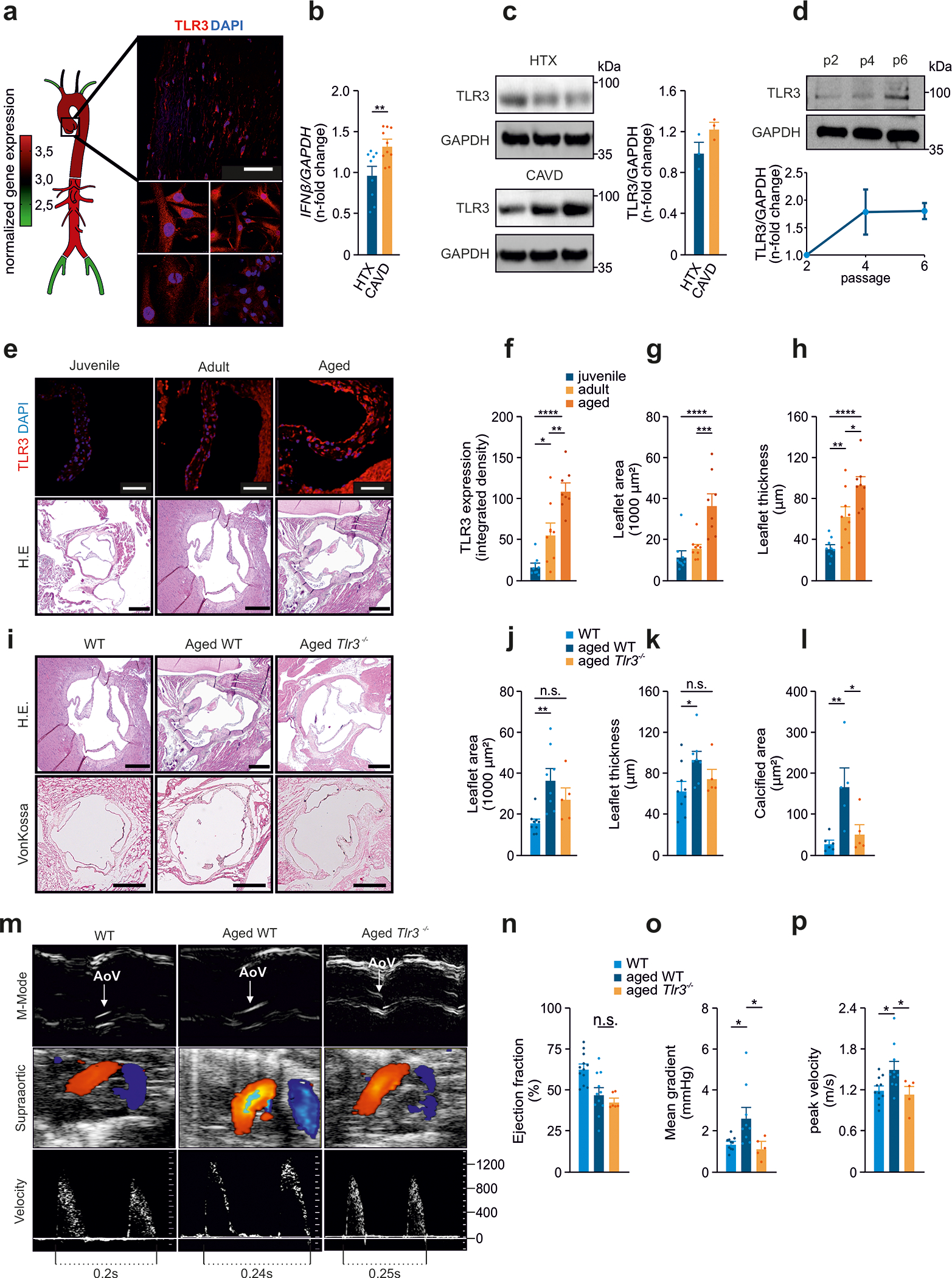
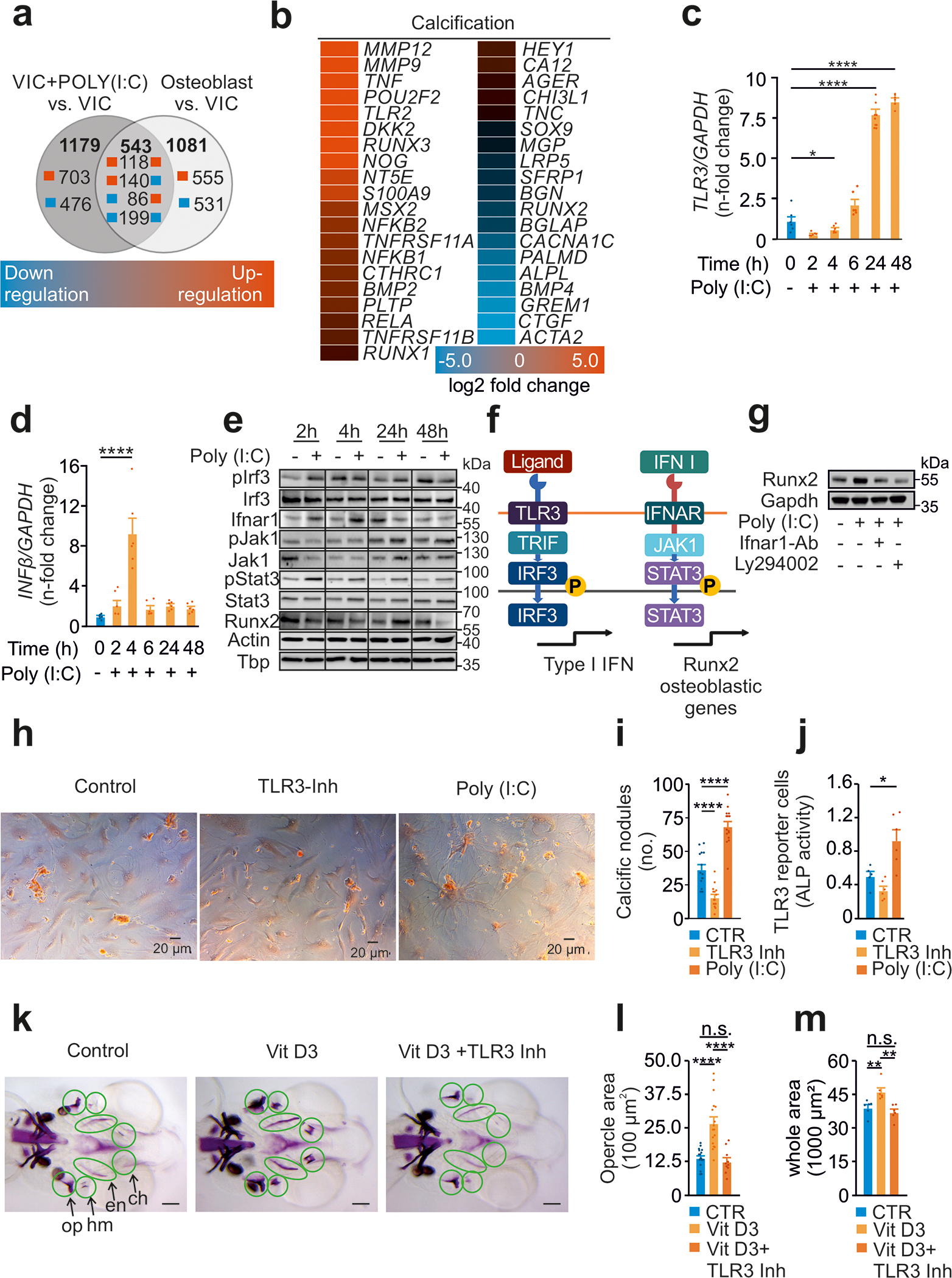
Background: Calcific aortic valve disease (CAVD) is characterized by a phenotypic switch of valvular interstitial cells to bone-forming cells. Toll-like receptors (TLRs) are evolutionarily conserved pattern recognition receptors at the interface between innate immunity and tissue repair. Type I interferons (IFNs) are not only crucial for an adequate antiviral response but also implicated in bone formation. We hypothesized that the accumulation of endogenous TLR3 ligands in the valvular leaflets may promote the generation of osteoblast-like cells through enhanced type I IFN signaling.

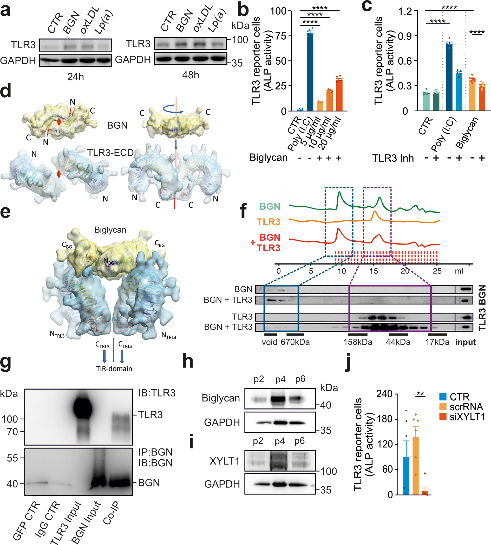
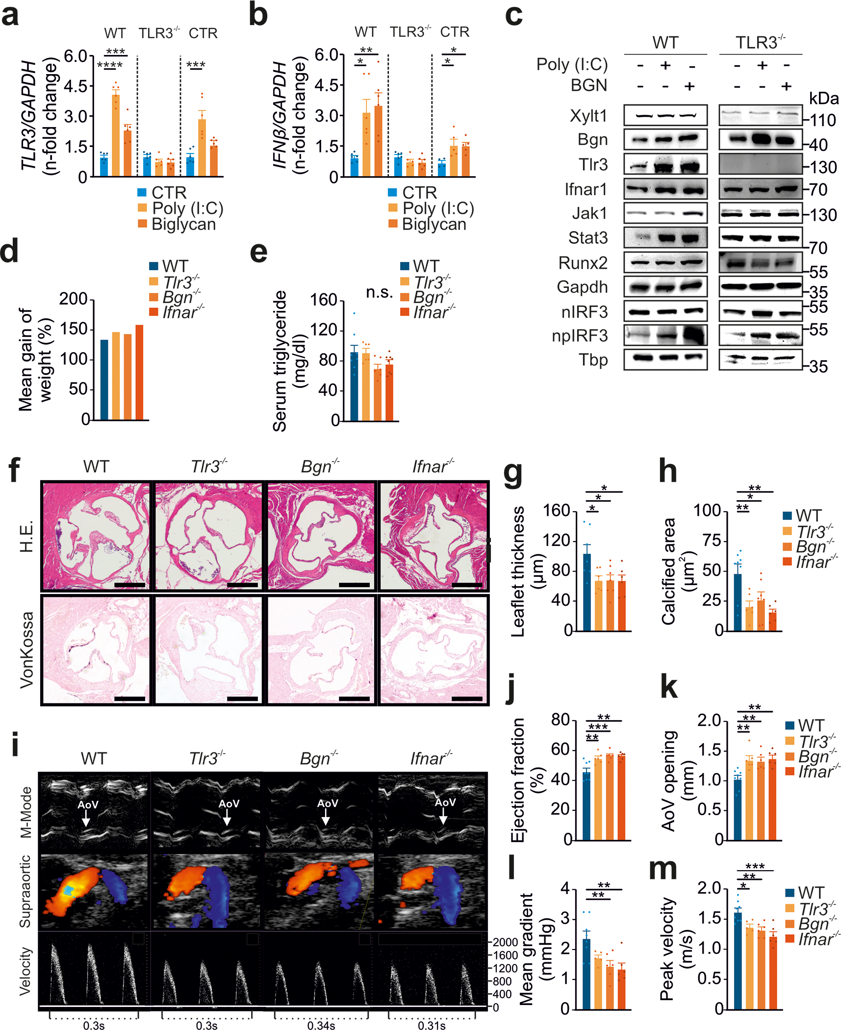
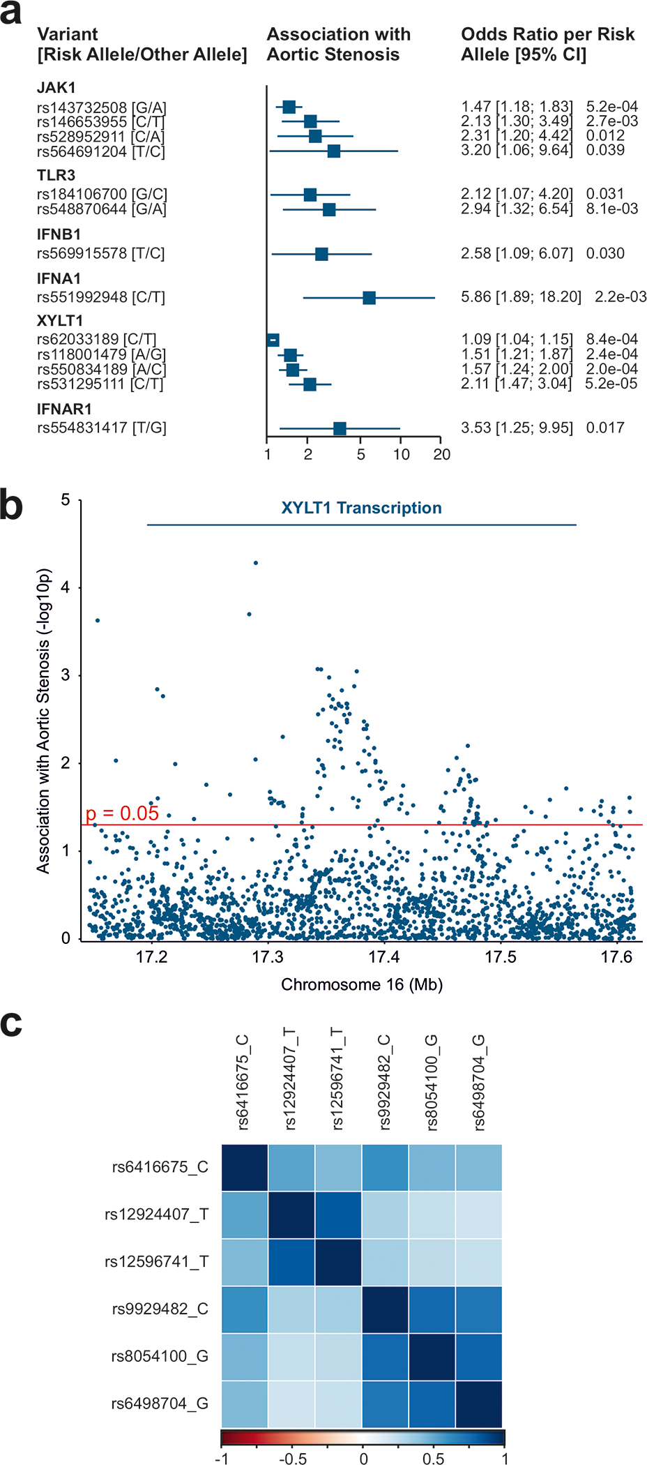
Methods: Human valvular interstitial cells isolated from aortic valves were challenged with mechanical strain or synthetic TLR3 agonists and analyzed for bone formation, gene expression profiles, and IFN signaling pathways. Different inhibitors were used to delineate the engaged signaling pathways. Moreover, we screened a variety of potential lipids and proteoglycans known to accumulate in CAVD lesions as potential TLR3 ligands. Ligand-receptor interactions were characterized by in silico modeling and verified through immunoprecipitation experiments. Biglycan (Bgn), Tlr3, and IFN-α/β receptor alpha chain (Ifnar1)-deficient mice and a specific zebrafish model were used to study the implication of the biglycan (BGN)-TLR3-IFN axis in both CAVD and bone formation in vivo. Two large-scale cohorts (GERA [Genetic Epidemiology Research on Adult Health and Aging], n=55 192 with 3469 aortic stenosis cases; UK Biobank, n=257 231 with 2213 aortic stenosis cases) were examined for genetic variation at genes implicated in BGN-TLR3-IFN signaling associating with CAVD in humans.

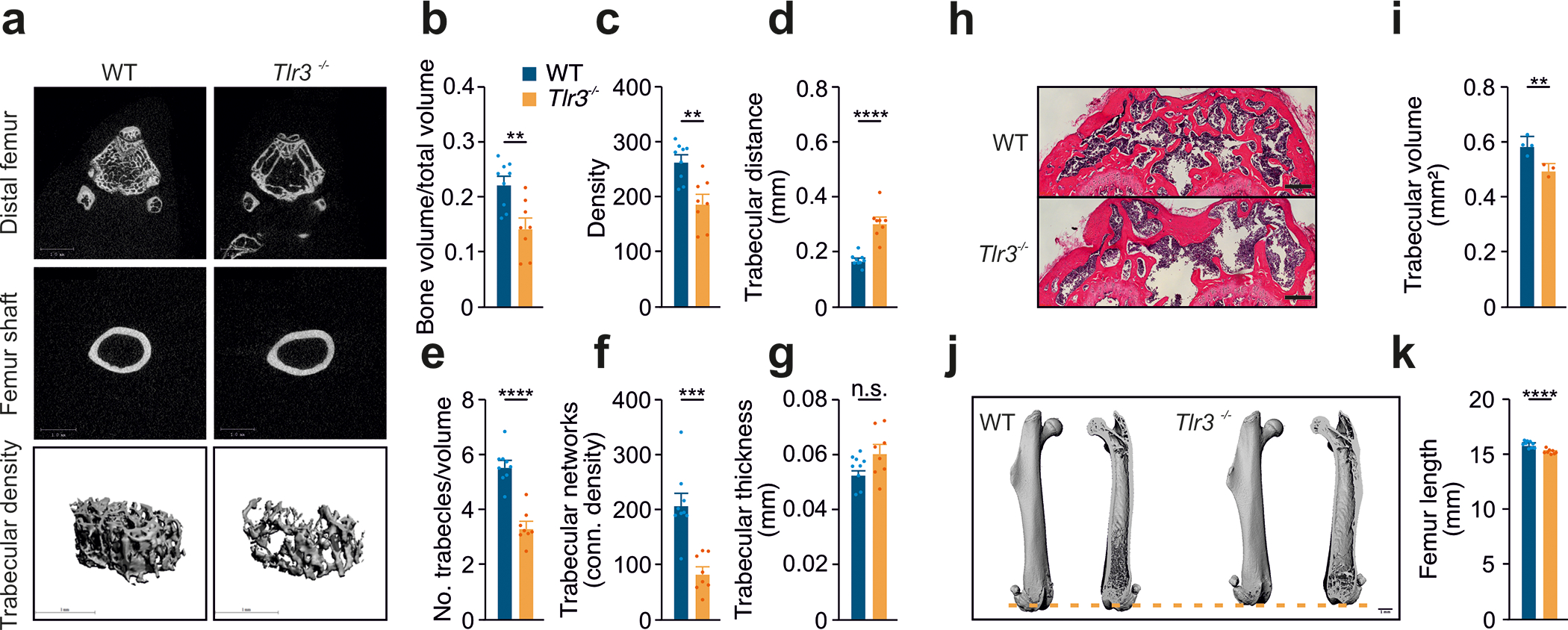
Results: Here, we identify TLR3 as a central molecular regulator of calcification in valvular interstitial cells and unravel BGN as a new endogenous agonist of TLR3. Posttranslational BGN maturation by xylosyltransferase 1 (XYLT1) is required for TLR3 activation. Moreover, BGN induces the transdifferentiation of valvular interstitial cells into bone-forming osteoblasts through the TLR3-dependent induction of type I IFNs. It is intriguing that Bgn-/-, Tlr3-/-, and Ifnar1-/- mice are protected against CAVD and display impaired bone formation. Meta-analysis of 2 large-scale cohorts with >300 000 individuals reveals that genetic variation at loci relevant to the XYLT1-BGN-TLR3-interferon-α/β receptor alpha chain (IFNAR) 1 pathway is associated with CAVD in humans.

Conclusions: This study identifies the BGN-TLR3-IFNAR1 axis as an evolutionarily conserved pathway governing calcification of the aortic valve and reveals a potential therapeutic target to prevent CAVD.

Keywords: Toll-like receptor 3; aortic valve disease; biglycan; extracellular matrix; osteogenesis; proteins.


Conflict of interest statement

Disclosures None.

Figures

Figure 1:
Figure 1:. TLR3 deficiency protects against age-related CAVD
(a) Analysis of Tlr3 expression in murine ascending aortas, descending aortas, subclavian arteries, carotid arteries and femoral arteries. Analysis of TLR3 levels in healthy human aortic valves (upper panel) and valvular interstitial cells from human aortic valves (lower panel); red: TLR3, blue: DAPI, scale=50 μm; n=3. (b) IFN-β expression, assessed by RT-PCR for CAVD samples and controls; n=9–10 per group. (c) Immunoblot analysis of TLR3 levels in human aortic valve specimens from CAVD patients and healthy controls; n=3. (d) Increasing levels of TLR3 with increasing passage number, for VICs analyzed by immunoblotting; n=2 independent western blotting experiments, representative western blot shown. (e) Representative images of murine aortic valves from juvenile (3 weeks), adult (12 weeks) and aged (>18 months) mice (red: TLR3, blue: DAPI, IF-scale=50 μm, H.E.-scale=250 μm) analyzed for (f) Tlr3 expression (n=8–9) and (g) leaflet area (n=8–9) and (h) leaflet thickness (n=8–9). (i) Aortic valves from adult wild-type mice (12 weeks), aged wild-type mice (>18 months) and aged Tlr3−/− mice (>18 months) analyzed by H.E staining for morphological analysis, and by vonKossa staining. (j) Aortic valve leaflet area measured on H.E. sections (n=5–9). (k) Aortic valve leaflet thickness measured on H.E. sections (n=5–9). (l) Calcified area of the aortic valve leaflets analyzed by vonKossa staining (n=5–6). (m) Echocardiographic assessment of aortic valves (AoV) from adult wild-type mice, aged wild-type mice and aged Tlr3−/− mice for the measurement of: (n) Ejection fraction (n=8–12) (o) Mean pressure gradients (n=8–12) and (p) Mean transvalvular velocity (n=8–12). All data are presented as the mean ± SEM. Statistical comparisons: (b) Two-tailed unpaired t-test, **P<0.01. (f-p) One-way ANOVA with Tukey post hoc tests: n.s. = non-significant, *P<0.05, **P<0.01, ***P<0.001, ****P<0.0001.
Figure 2:
Figure 2:. TLR3 is a conserved inducer of calcification via IFN I signaling.
(a) Numbers of at least four-fold change differentially expressed genes in human VICs with or without Poly(I:C) treatment (20 μg/mL) for 72h and human osteoblasts. (b) Expression profile of genes involved in calcification. (c) RT-PCR analysis of VICs treated with Poly(I:C) for TLR3 (n=6) and (d) IFN-β (n=6), measured in duplicates. (e) Immunoblot analysis of VICs treated with Poly(I:C) assessing the TLR3-RUNX2 pathway (n=2 independent western blot experiments, representative western blot shown). (f) Schematic illustration of the involved TLR3-RUNX2 pathway. (g) Expression of the osteoblastic transcription factor RUNX2 after Poly(I:C) treatment (20 μg/mL) in the presence of either LY294002 (10μM), an IRF3 inhibitor, or a specific IFNAR1 blocking antibody (4μg/ml). GAPDH served as loading control (n=2 independent western blot experiments, representative western blot shown). (h) VICs cultured with osteoblastic medium in the presence of TLR3 agonist Poly(I:C) or inhibitor (scale=40μm). (i) Quantification of calcific nodules upon treatment applying Alizarin Red staining (n=9). (j) Activity of alkaline phosphatase measured in the supernatant of treated VICs (n=9). (k) Zebrafish treated with VitD3 in the presence of TLR3 inhibitor with subsequent quantification of calcification of (l) the opercle (n=10–19, scale=100μm) (m) all bone structures (n=6, scale=100μm). All data are presented as mean ± SEM. Statistical comparisons: One-way ANOVA with Tukey post hoc test: *P<0.05, **P<0.01, ****P<0.0001
Figure 3:
Figure 3:. Tlr3−/− exhibit osteoporotic phenotype.
(a) Femurs from 12-week-old wild type and Tlr3−/− (n=8–9 femora) analyzed morphologically via micro-CT for (b) bone volume, (c) bone density, (d) trabecular distance, (e) number of trabecles/volume, (f) trabecular networks and (g) trabecular thickness. (h) Histological analysis of distal femurs from 12-week-old wild type and Tlr3−/− analyzing (i) trabecular volume (H.E. staining, n=3–4). (j,k) Femur length from 12-week-old wild type and Tlr3−/− analyzed via micro-CT (n=8–9 femora). All data are presented as mean ± SEM. Statistical comparisons: 2-tailed unpaired t-test, *P<0.05, **P<0.01, ***P<0.001, ****P<0.0001.
Figure 4:
Figure 4:. Biglycan serves as an endogenous ligand of TLR3.
(a) Immunoblot analysis of human VICs treated with biglycan (20 μg/mL), Lp(a) (20 μg/mL) or oxLDL (20 μg/mL) for 24h and 48h for TLR3 protein expression. (b) HEK293 hTLR3 reporter cells treated with Poly(I:C) (20 μg/ml) and various concentrations of biglycan (5 μg/ml, 10 μg/ml, 20 μg/ml), (n=6). (c) HEK293 hTLR3 reporter cells treated with Poly(I:C) or biglycan (both at 5 μg/ml) for 6 h in the presence of (R)-2-(3-chloro-6-fluorobenzo[b]thiophene-2-carboxamido)-3-phenylpropanoic acid, a dsRNA/TLR3 complex inhibitor (n=6). (d) Illustration of the dimer binding mode. The top view (left panel) shows the location of the 2-fold axes perpendicular to the projection plane with dyad symbols, and the right panel shows the assembly of the model maintaining the 2-fold symmetry (red line). The docked, refined, and optimized model (e) is presented as ribbons surrounded by a macro shape which includes the forests core residues of all glycan decorations, none of which cause any steric clashes. (f) Binding experiments with the recombinant human TLR3 ectodomain and biglycan (size-exclusion chromatography and subsequent immunoblot analysis). (g) Following the co-immunoprecipitation of purified TLR3 ectodomain and purified BGN, both proteins were detected on immunoblots (n=2 independent experiments, representative blots shown). (h,i) Increasing levels of BGN and XYLT1 in VICs with increasing passage number analyzed by immunoblotting (n=2 independent western blotting experiments, representative western blot shown) (j) HEK293 hTLR3 reporter cells treated with supernatants from VICs after siRNA knockdown of XYLT1 or after treatment with a control RNA (scRNA) (n=6). Statistical comparisons: (b-c,k) One-way ANOVA with Tukey post hoc test: **P<0.01, ****P<0.0001.
Figure 5:
Figure 5:. The BGN-TLR3-IFNAR1 axis induces aortic valve calcification
(a) RT-PCR analysis of human wild-type dermal fibroblasts, TLR3−/− CRISPR fibroblasts or empty vector-transformed control fibroblasts (CTR) treated with Poly(I:C) or BGN (both at 5 μg/ml) for 6h and then subjected to an analysis of TLR3 expression (n=6). (b) RT-PCR analysis of human wild-type dermal fibroblasts, TLR3−/− CRISPR fibroblasts or empty vector-transformed control fibroblasts (CTR) were treated with Poly(I:C) or BGN (both at 5 μg/ml) for 6 h and then analyzed for IFN-ß expression (n=6). (c) Immunoblot analysis of human wild-type dermal fibroblasts and TLR3−/− CRISPR fibroblasts treated with Poly(I:C) or BGN (both at 5 μg/ml) for 6h. (d) Mean weight gain after four months on a high-fat diet, for wild-type, Tlr3−/−, Bgn−/− and Ifnar1−/− animals (n=6–8). (e) Serum triglyceride concentration after 4 months on a high-fat diet, for wild-type, Tlr3−/−, Bgn−/− and Ifnar1−/− animals (n=6–8 animals per group). (f) Histological evaluation of aortic valves from wild-type, Tlr3−/−, Bgn−/− or Ifnar1−/− animals fed a high-fat diet for four weeks by H.E. and vonKossa staining. (g) Analysis of aortic valve leaflet thickness in wild-type, Tlr3−/−, Bgn−/− and Ifnar1−/− animals after 4 months on a high-fat diet (n=6–8). (h) Quantification of aortic valve calcification in wild-type, Tlr3−/−, Bgn−/− and Ifnar1−/− animals after 4 months on a high-fat diet (n=6–8). (i) Assessment, by transthoracic echocardiography, of aortic valve function after 4 months on a high-fat diet, for wild-type, Tlr3−/−, Bgn−/− and Ifnar1−/− animals (n=6–8). (j) Analysis of ejection fraction in wild-type, Tlr3−/−, Bgn−/− and Ifnar1−/− animals after 4 months on a high-fat diet (n=6–8). (k) Aortic valve opening in wild-type, Tlr3−/−, Bgn−/− and Ifnar1−/− animals after 4 months on a high-fat diet (n=6–8). (l) Mean transvalvular pressure gradient in wild-type, Tlr3−/−, Bgn−/− and Ifnar1−/− animals after 4 months on a high-fat diet (n=5–8). (m) Transvalvular peak velocities in wild-type, Tlr3−/−, Bgn−/− and Ifnar1−/− animals after 4 months on a high-fat diet (n=5–8). All data are presented as the mean ± SEM. Statistical comparisons: (a), (b), (e), (g), (h), (j), (k), (l), (m) One-way ANOVA with Dunnett post hoc tests: *P<0.05, **P<0.01, ***P<0.05, ****P<0.000
Figure 6:
Figure 6:. Association of TLR3 pathway variants in aortic stenosis patients.
(a) Association with aortic stenosis (meta-analysis of UK Biobank and GERA) of variants within 50 kb of 6 autosomal genes. Variants shown are independent (r2 < 0.01) and have an association of 1) p ≤ 1 × 10⁻³ or 2) p ≤ 0.05 and odds ratio ≥ 2. (See Table 1 for a complete list of significant variants.) (b) Association with aortic stenosis (meta-analysis of UK Biobank and GERA) of variants within 50 kb of the XYLT1 transcript (Manhattan plot). (c) Correlation of variants (r2) in the UK Biobank participants of variants associated with XYLT1 expression.

References

    1. Danielsen R, Aspelund T, Harris TB, Gudnason V. The prevalence of aortic stenosis in the elderly in Iceland and predictions for the coming decades: the AGES-Reykjavik study. Int J Cardiol. 2014;176:916–922. doi: 10.1016/j.ijcard.2014.08.053 - DOI - PMC - PubMed
    1. Lindman BR, Clavel MA, Mathieu P, Iung B, Lancellotti P, Otto CM, Pibarot P. Calcific aortic stenosis. Nat Rev Dis Primers. 2016;2:16006. doi: 10.1038/nrdp.2016.6 - DOI - PMC - PubMed
    1. Rajamannan NM, Evans FJ, Aikawa E, Grande-Allen KJ, Demer LL, Heistad DD, Simmons CA, Masters KS, Mathieu P, O’Brien KD, et al. Calcific aortic valve disease: not simply a degenerative process: A review and agenda for research from the National Heart and Lung and Blood Institute Aortic Stenosis Working Group. Executive summary: Calcific aortic valve disease-2011 update. Circulation. 2011;124:1783–1791. doi: 10.1161/CIRCULATIONAHA.110.006767 - DOI - PMC - PubMed
    1. Liu AC, Joag VR, Gotlieb AI. The emerging role of valve interstitial cell phenotypes in regulating heart valve pathobiology. Am J Pathol. 2007;171:1407–1418. doi: 10.2353/ajpath.2007.070251 - DOI - PMC - PubMed
    1. Driscoll K, Cruz AD, Butcher JT. Inflammatory and Biomechanical Drivers of Endothelial-Interstitial Interactions in Calcific Aortic Valve Disease. Circ Res. 2021;128:1344–1370. doi: 10.1161/CIRCRESAHA.121.318011 - DOI - PMC - PubMed

Publication types

Supplementary concepts