Review
. 2023 May;16(5):173-181.
doi: 10.1007/s12178-023-09834-2. Epub 2023 Apr 4.

Ramp Lesions of the Medial Meniscus

Affiliations
Review

Ramp Lesions of the Medial Meniscus

Renaud Siboni et al. Curr Rev Musculoskelet Med. 2023 May.

Abstract

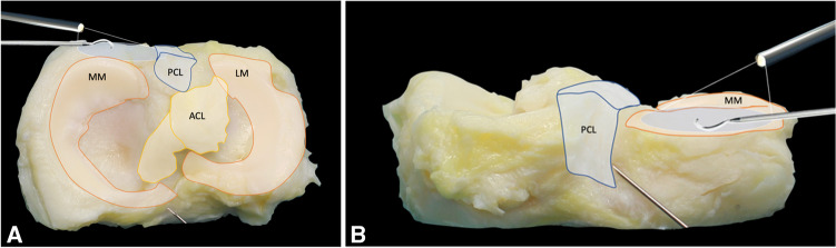
Purpose of review: To provide an overview of the recent scientific literature about ramp lesions of the medial meniscus and to summarise the current evidence on their prevalence, classification, biomechanics, surgical techniques and clinical outcomes.

Recent findings: Ramp lesions may be present in more than 1 patient undergoing ACL reconstruction out of 5 and almost half of the medial meniscal tears observed in this population. Due to the risk of persistent anterior and rotational laxity after ACL reconstruction, their repair has been advocated. There is no general agreement to date on whether and when ramp lesions should be treated surgically. Comparative studies have failed to show that the repair of stable lesions was superior in comparison to nonoperative approaches. A lower failure rate and secondary meniscectomy has been reported with a suture hook repair through the posteromedial portal in comparison with an all-inside technique. Furthermore, reconstructions of the anterolateral complex in association with ACL reconstruction may have a protective effect on ramp repair. Ramp lesions of the medial meniscus in ACL-injured knees cannot be neglected anymore. Given their novelty, their clinical impact has not been fully assessed yet, but the evidence is growing that they need to be systematically identified and eventually repaired, for which they require advanced surgical knowledge. There is, to date, no consensus on whether and when ramp lesions should be treated surgically. Their subtypes, size and stability may influence the decision-making process.

Keywords: ACL; Medial meniscus; Outcomes; Prevalence; Ramp lesion; Repair.


Conflict of interest statement

The authors declare no competing interests.

Figures

Fig. 1
Fig. 1
Magnetic resonance imaging (MRI) of a ramp lesion of the medial meniscus. Sagittal slice MRI of a typical sign of a ramp lesion in an ACL-deficient knee. In this right knee, a hyper T2-weighted signal can be observed at the meniscocapsular junction of the medial meniscus posterior horn. Abbreviations: MFC: medial femoral condyle; MTP: medial tibial plateau; ACL: anterior cruciate ligament; MCJ, meniscocapsular junction; MM: medial meniscus
Fig. 2
Fig. 2
Arthroscopic classification of ramp lesions according to Thaunat et al. [29]. Five different types of ramp lesions are distinguished during arthroscopy: type 1: meniscocapsular lesions; type 2: partial superior lesions; type 3: partial inferior or hidden lesions; type 4: complete tears in the red-red zone; type 5: double tears. Abbreviations: MFC: medial femoral condyle; MTP: medial tibial plateau
Fig. 3
Fig. 3
Magnetic resonance imaging (MRI) classification of ramp lesions according to Greif et al. [••]. Five different types of ramp lesions are distinguished on MRI: type 1: meniscocapsular ligament tear; type 2: partial superior peripheral meniscal horn tear; type 3A: partial inferior peripheral posterior horn meniscal tear; type 3B: meniscotibial ligament tear; type 4A: complete peripheral posterior horn meniscal tear; type 4B: complete meniscojunction tear; type 5: peripheral posterior horn meniscal double tear. Abbreviations: MFC: medial femoral condyle; MTP: medial tibial plateau
Fig. 4
Fig. 4
All-inside suture hook through a posteromedial approach with a trans-condylar notch arthroscopic control. Superior (A) and posterior (B) views of an anatomical section of the upper end of the tibia. Illustration of how the arthroscope is situated through the notch, between the posterior cruciate ligament and the medial femoral condyle. The suture hook is inserted through a posteromedial approach. Abbreviations: MM: medial meniscus; LM: lateral meniscus; PCL: posterior cruciate ligament; ACL: anterior cruciate ligament
Fig. 5
Fig. 5
All-inside suture hook through a 2-portal posteromedial approach. Superior (A) and posterior (B) views of an anatomical section of the upper end of the tibia. Placement of the arthroscope through a first posteromedial portal and the suture hook through a second posteromedial approach. Abbreviations: MM: medial meniscus; LM: lateral meniscus; PCL: posterior cruciate ligament; ACL: anterior cruciate ligament
Fig. 6
Fig. 6
All-inside suture hook through a 2-portal posteromedial approach – arthroscopic views. This is a right knee at 90° knee flexion. (A) The entry of the posteromedial viewing portal is identified through a trans-notch articular view with the help of a needle and trans-illumination to protect from saphenous vessel injury. (B) Direct visualisation of the posteromedial corner and a ramp lesion. The camera is in the posteromedial viewing portal. (C) The lesion is debrided with a shaver through the second posteromedial portal located approximately 3 to 4 cm posteriorly and distally to the viewing portal. (D). Suture of a ramp lesion with a 90° curved hook and a PDS 1 wire. (E) After suture retrieval, a sliding knot is made using a knot pusher. (F) After section of the suture, the knot can be visualised. Abbreviations: PMC: posteromedial capsule; MM: medial meniscus; MFC: Medial femoral condyle; MCJ: meniscocapsular junction
Fig. 7
Fig. 7
Posteromedial view before and after repair of a ramp lesion and during flexion and extension. This is a right knee. The camera is in the posteromedial viewing portal. Before repair, the ramp lesion is observed at 90° (A) and 20° (B) knee flexion. A cleft between the posterior wall of the medial meniscus and the ramp tissue can be identified in both positions. After repair, posteromedial view at 90° (C) and 20°(D) knee flexion. The black star indicates adequate tensioning of the posterior capsule by the repair (B and D). Abbreviations: MFC: medial femoral condyle; MM: medial meniscus; MCJ: meniscocapsular junction; PMC: posteromedial capsule

References

    1. Hamberg P, Gillquist J, Lysholm J. Suture of new and old peripheral meniscus tears. J Bone Joint Surg Am. 1983;65(2):193–197. doi: 10.2106/00004623-198365020-00007. - DOI - PubMed
    1. Lemaire M, Combelles F, Miremad C, Van Vooren P. Postero-internal menisco-capsular disinsertions associated with chronic instabilities of the knee caused by rupture of the anterior cruciate ligament. Rev Chir Orthop Reparatrice Appar Mot. 1984;70(8):613–622. - PubMed
    1. Ahn JH, Kim SH, Yoo JC, Wang JH. All-inside suture technique using two posteromedial portals in a medial meniscus posterior horn tear. Arthroscopy. 2004;20(1):101–108. doi: 10.1016/j.arthro.2003.11.008. - DOI - PubMed
    1. Ahn JH, Wang JH, Yoo JC. Arthroscopic all-inside suture repair of medial meniscus lesion in anterior cruciate ligament–deficient knees: results of second-look arthroscopies in 39 cases. Arthroscopy. 2004;20(9):936–945. doi: 10.1016/j.arthro.2004.06.038. - DOI - PubMed
    1. Arnoczky SP, Warren RF. Microvasculature of the human meniscus. Am J Sports Med. 1982;10(2):90–95. doi: 10.1177/036354658201000205. - DOI - PubMed