. 2023 May 8;8(9):e161118.
doi: 10.1172/jci.insight.161118.

RBM47 regulates intestinal injury and tumorigenesis by modifying proliferation, oxidative response, and inflammatory pathways

Affiliations

RBM47 regulates intestinal injury and tumorigenesis by modifying proliferation, oxidative response, and inflammatory pathways

Saeed Soleymanjahi et al. JCI Insight. .

Abstract

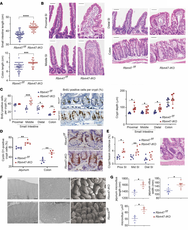
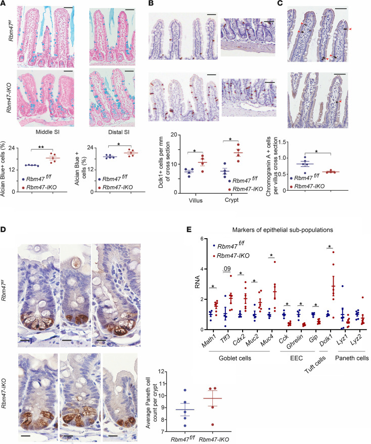
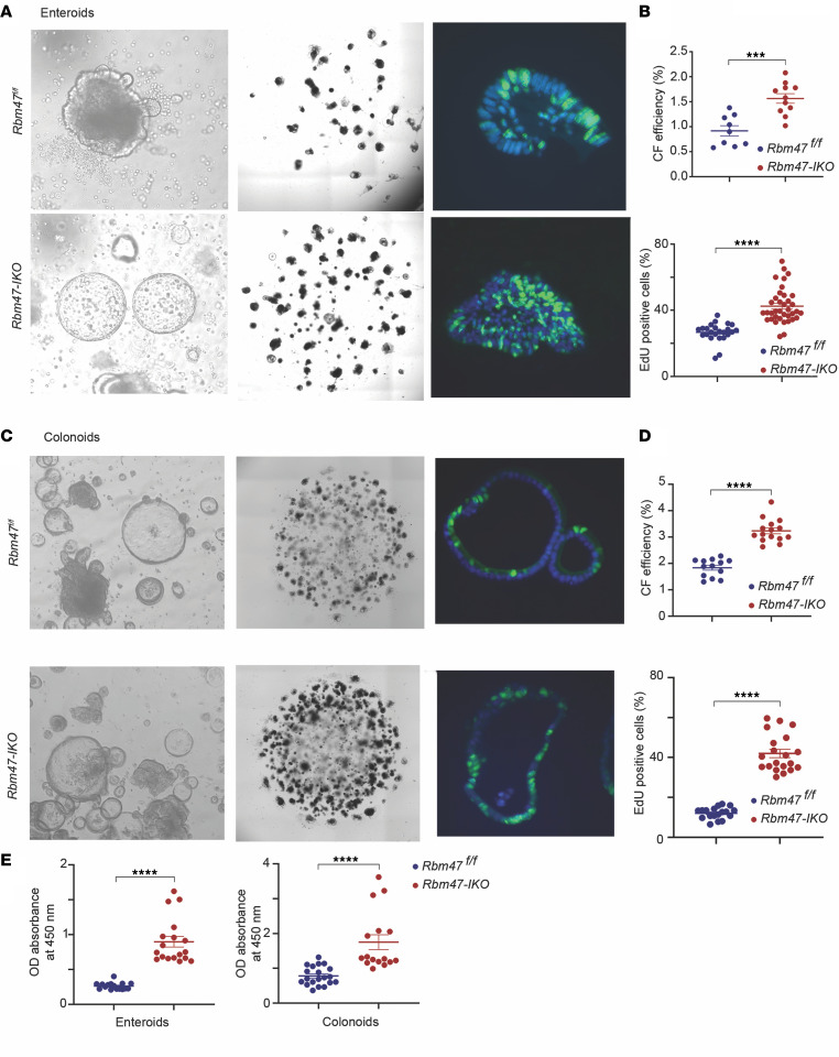
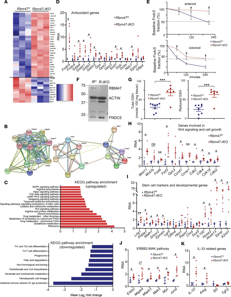
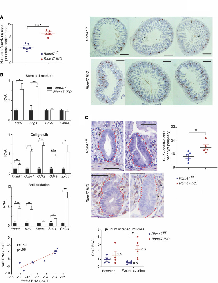
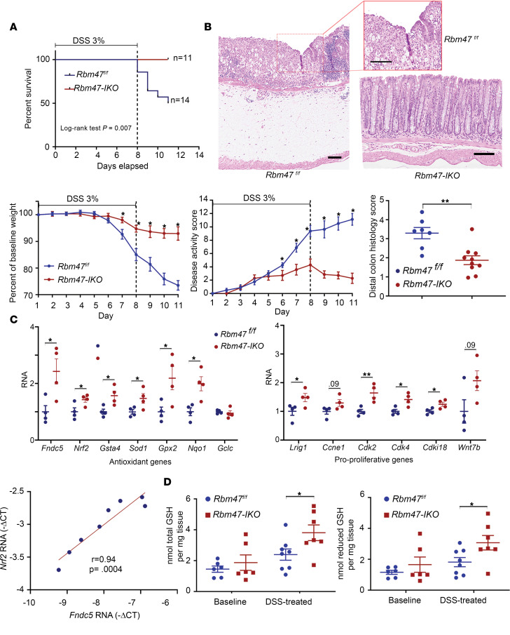
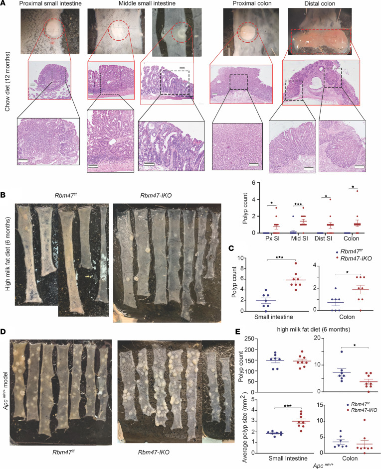
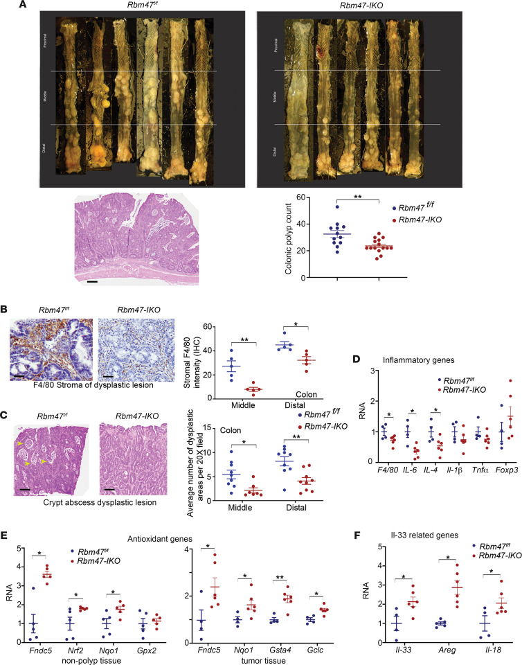
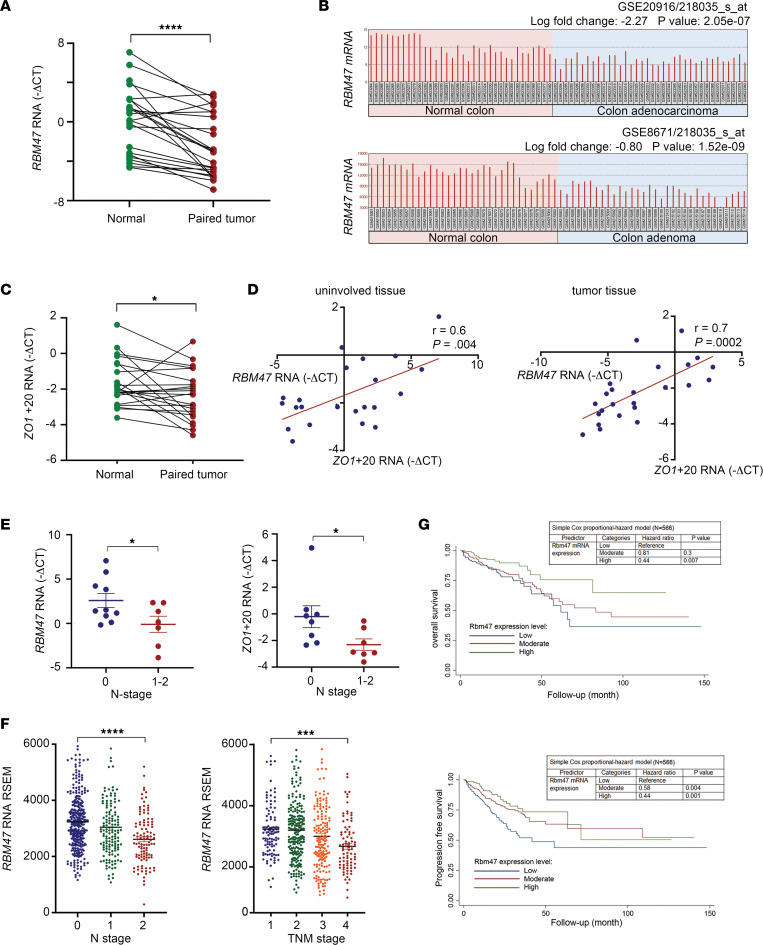
RNA-binding protein 47 (RBM47) is required for embryonic endoderm development, but a role in adult intestine is unknown. We studied intestine-specific Rbm47-knockout mice (Rbm47-IKO) following intestinal injury and made crosses into ApcMin/+ mice to examine alterations in intestinal proliferation, response to injury, and tumorigenesis. We also interrogated human colorectal polyps and colon carcinoma tissue. Rbm47-IKO mice exhibited increased proliferation and abnormal villus morphology and cellularity, with corresponding changes in Rbm47-IKO organoids. Rbm47-IKO mice adapted to radiation injury and were protected against chemical-induced colitis, with Rbm47-IKO intestine showing upregulation of antioxidant and Wnt signaling pathways as well as stem cell and developmental genes. Furthermore, Rbm47-IKO mice were protected against colitis-associated cancer. By contrast, aged Rbm47-IKO mice developed spontaneous polyposis, and Rbm47-IKO ApcMin/+ mice manifested an increased intestinal polyp burden. RBM47 mRNA was decreased in human colorectal cancer versus paired normal tissue, along with alternative splicing of tight junction protein 1 mRNA. Public databases revealed stage-specific reduction in RBM47 expression in colorectal cancer associated independently with decreased overall survival. These findings implicate RBM47 as a cell-intrinsic modifier of intestinal growth, inflammatory, and tumorigenic pathways.

Keywords: Colorectal cancer; Gastroenterology; Inflammation; RNA processing.


Conflict of interest statement

Conflict of interest: The authors have declared that no conflict of interest exists.

Figures

Figure 1
Figure 1. Constitutive intestinal Rbm47 deletion enhances the proliferation capacity and anatomical development of intestinal epithelium in young (8–14 weeks) mice.
(A) Small intestine (top) and colon (bottom) length in Rbm47fl/fl and Rbm47-IKO mice (unpaired t test, n = 55 fl/fl, 37 IKO for small intestine and 43 fl/fl, 24 IKO for colon). (B) Top: regional sections of small intestine and colon from Rbm47fl/fl and Rbm47-IKO mice (scale bar: 50 μm). Bottom: Crypt depth in Rbm47fl/fl and Rbm47-IKO mice (unpaired t test, n = 6/genotype, 30–40 crypts with longitudinal cross-sectional view per mouse). (C) Left: Regional BrdU-positive abundance (% per total crypt cells) in small intestine and colon from Rbm47fl/fl and Rbm47-IKO mice (unpaired t test, n = 6/genotype, 20–30 crypts with complete longitudinal cross-sectional view per mouse). Right: BrdU-stained sections of middle small intestine crypts (scale bar: 25 μm). (D) Left: Cyclin D1–positive abundance (% per total crypt cells) in jejunum and colon from young Rbm47fl/fl and Rbm47-IKO mice (unpaired t test, n = 5/genotype, 20–30 crypts with longitudinal cross-sectional view per mouse); Right: Cyclin D1-stained sections of middle small intestine crypts (scale bar: 25 μm). (E) Left: regional crypt fission in small intestine from Rbm47fl/fl and Rbm47-IKO mice (unpaired t test, n = 6/genotype, 200 crypts with longitudinal cross-sectional view per mouse per section). Right: H&E-stained view of crypt fission (scale bar: 50 μm). (F) Transmission electron microscopic sections exhibiting length (left) and cross-sectional density (middle) of microvilli (scale bar: 500 nm) and scanning electron microscopic sections of villi (right, scale bar: 100 μm) in middle small intestine of young mice. (G) Left: Length (top) and density (bottom) of microvilli in middle small intestine from Rbm47fl/fl and Rbm47-IKO mice (unpaired t test, n = 3/genotype, 75–100 microvilli per mouse). Right: middle small intestine villi width in Rbm47fl/fl and Rbm47-IKO mice (unpaired t test, n = 6/genotype, 60–80 villi per mouse). Data are presented as mean ± SEM. *P < 0.05, **P < 0.01, ***P < 0.001, and ****P < 0.0001.
Figure 2
Figure 2. Constitutive intestinal Rbm47 deletion alters abundance of differentiated cell subpopulations in intestinal epithelium of young (8–14 weeks) mice.
(A) Top: Alcian blue–stained sections of middle (left) and distal (right) small intestine in Rbm47fl/fl and Rbm47-IKO mice (scale bar: 25 μm). Bottom: goblet cell abundance (% per total cells) in villi and crypts of middle (left graph) and distal (right graph) small intestine from Rbm47fl/fl and Rbm47-IKO mice (unpaired t test, n = 5/genotype, 30–40 villi and crypts with complete longitudinal cross-sectional view per mouse). (B) Top: Dclk1-stained sections of middle small intestine villi (left) and crypts (right) to demonstrate tuft cells in Rbm47fl/fl and Rbm47-IKO mice (scale bar: 25 μm). Bottom: tuft cell abundance (cell count/mm of border) in villi and crypts of middle small intestine from Rbm47fl/fl and Rbm47-IKO mice (unpaired t test, n = 4/genotype, 30–40 villi and crypts with complete longitudinal cross-sectional view per mouse). (C) Top: Chromogranin A–stained sections of middle small intestine villus to demonstrate enteroendocrine cells (EEC) (red arrowheads) in Rbm47fl/fl and Rbm47-IKO mice (scale bar: 25 μm). Bottom: EEC abundance (cell count per longitudinal cross-section of villus) in middle small intestine from Rbm47fl/fl and Rbm47-IKO mice (unpaired t test, n = 5 fl/fl and 4 IKO, 30–40 villi with complete longitudinal cross-sectional view per mouse). (D) Left: Lysozyme-stained sections of middle small intestine crypts to demonstrate Paneth cells in Rbm47fl/fl and Rbm47-IKO mice (scale bar: 20 μm). Right: Paneth cell abundance (cell count per crypt) in middle small intestine from Rbm47fl/fl and Rbm47-IKO mice (unpaired t test, n = 5/genotype, 35–40 crypts with complete longitudinal cross-sectional view per mouse). (E) qPCR evaluation of different epithelial subpopulation cell markers in middle small intestine from young Rbm47fl/fl and Rbm47-IKO mice (unpaired t test, n = 4 fl/fl, 6 IKO). Data are presented as mean ± SEM; *P < 0.05, and **P < 0.01.
Figure 3
Figure 3. Constitutive intestinal Rbm47 deletion increases cell viability and proliferation in young (8–14 weeks) mice’s stem cell–derived enteroids and colonoids.
(A) Left: inverse light microscopic view of Rbm47fl/fl and Rbm47-IKO mice’s middle small intestine stem cell–derived organoids. Middle: Cytation 5 scanner view of colony-forming assay (CFA) using middle small intestine stem cell–derived organoids. Right: fluorescence microscopic images of EdU-stained (shown in green) middle small intestine stem cell–derived organoids. Hoechst (blue) was used for nuclear staining. (B) CFA (top) and abundance of EdU-positive cells (% of total cells per organoid) (bottom) in Rbm47fl/fl and Rbm47-IKO mice’s middle small intestine stem cell–derived organoids (mean ± SEM, unpaired t test, 9 fl/fl and 11 IKO experimental replicates for CFA; 24 fl/fl and 37 IKO organoids for EdU assay); ***P < 0.001; ****P < 0.0001. (C) Left: inverse light microscopic view of Rbm47fl/fl and Rbm47-IKO mice stem cell–derived colonoids. Middle: Cytation 5 scanner view of CFA using the colonoids. Right: fluorescence microscopic images of EdU-stained (green) colonoids. (D) CFA (top) and abundance of EdU-positive cells (% of total cells per organoid) (bottom) in Rbm47fl/fl and Rbm47-IKO mice’s stem cell–derived colonoids (mean ± SEM, unpaired t test, 13 fl/fl and 15 IKO experimental replicates for CFA; 18 fl/fl and 20 IKO organoids for EdU assay); ****P < 0.0001. (E) Cell viability assay using cell counting kit-8 method in Rbm47fl/fl and Rbm47-IKO mice’s stem cell–derived enteroids (left) and colonoids (right) (mean ± SEM, unpaired t test; n = 16 fl/fl and 18 IKO experimental replicates for enteroids; n = 20 fl/fl and 16 IKO experimental replicates for colonoids); ****P < 0.0001.
Figure 4
Figure 4. Differentially expressed genes in small intestine from young (8–14 weeks) Rbm47-IKO mice.
(A) Heatmap illustration of top upregulated and downregulated genes in Rbm47-IKO versus Rbm47fl/fl mice. (B) STRING analysis of upregulated genes related to oxidative response (left) and WNT signaling (right). (C) Enriched Kyoto Encyclopedia of Genes and Genomes (KEGG) pathways in upregulated (top) and downregulated (bottom) genes. (D) qPCR evaluation of differentially upregulated genes involved in antioxidative response and glutathione metabolism (mean ± SEM, unpaired t test, n = 4 fl/fl and 6 IKO); *P < 0.05. (E) Relative Fndc5 mRNA abundance at successive time points after actinomycin D treatment as a fraction of baseline Fndc5 in enteroids (top) and colonoids (bottom) (mean ± SEM, unpaired t test, n = 3 independent experiments per genotype/time point); *P < 0.05. (F) Abundance of RBM47, ACTIN, and FNDC5 proteins in middle small intestine mucosa from Rbm47fl/fl and Rbm47-IKO mice (each lane represents a pool of 3 separate extracts per genotype). (G) Tissue content of total (left) and reduced (right) glutathione in middle small intestine from young mice (mean ± SEM, unpaired t test, n = 8 fl/fl and 6 IKO). ***P < 0.001. (HK) qPCR evaluation of differentially upregulated genes involved in WNT signaling and cell proliferation (H), stem cell markers and developmental genes (I), genes involved in ERBB2/MAPK pathway (J), and IL-33–related genes (K) (mean ± SEM, unpaired t test, n = 4 fl/fl and 6 IKO); *P < 0.05, **P < 0.01.
Figure 5
Figure 5. Constitutive intestinal Rbm47 deletion promotes recovery of small intestine after 12 Gy whole-body irradiation.
(A) Left: number of surviving crypts, determined using modified microcolony assay, per middle small intestine cross-sectional area in Rbm47fl/fl and Rbm47-IKO mice, 84 hours after whole-body irradiation (mean ± SEM, unpaired t test, n = 5/genotype, 6–8 cross sections per mouse). Right: BrdU-stained cross sections of middle small intestine from Rbm47fl/fl and Rbm47-IKO mice after irradiation (scale bar: 400 μm). (B) qPCR evaluation of stem cell markers, cell growth genes, and antioxidative genes’ expressions as well as the association between Fndc5 and Nrf2 expressions in middle small intestine from irradiated Rbm47fl/fl and Rbm47-IKO mice (mean ± SEM and unpaired t test for gene expressions, Pearson’s r test for correlation, n = 5/genotype); *P < 0.05, **P < 0.01, ***P < 0.001. (C) Top left: COX2 immunohistochemical staining of middle small intestine from irradiated Rbm47fl/fl and Rbm47-IKO mice (scale bar: 50 μm). Pericryptal COX2-positive cells are marked by black arrowheads. Top right: number of pericryptal COX2-positive cells/mm crypt periphery (mean ± SEM, unpaired t test, n = 5/genotype, 20–30 crypts with complete longitudinal cross-sectional view per mouse); *P < 0.05. Bottom: Cox2 mRNA expression in middle small intestine from Rbm47fl/fl and Rbm47-IKO mice before and after irradiation (mean ± SEM, unpaired t test, n = 4/genotype for each assay); *P < 0.05.
Figure 6
Figure 6. Constitutive intestinal Rbm47 deletion attenuates colitis after 3% DSS treatment.
(A) Survival rates (top), percentages of baseline body weight loss (bottom left), and disease activity scores (bottom right) of male Rbm47fl/fl and Rbm47-IKO mice treated with 3% DSS (unpaired t test, n = 14 fl/fl and 11 IKO); *P < 0.05. (B) Representative H&E-stained sections (top, scale bar: 100 μm) and microscopic histology score (bottom) of distal colon from Rbm47fl/fl and Rbm47-IKO mice treated with 3% DSS (mean ± SEM, unpaired t test, n = 7 fl/fl and 10 IKO); **P < 0.01. (C) qPCR evaluation of genes involved in antioxidative response and glutathione metabolism (top left), stem cell markers and cell proliferation genes (top right), and association between Fndc5 and Nrf2 expressions (bottom) in colons from DSS-treated male Rbm47fl/fl and Rbm47-IKO mice (mean ± SEM and unpaired t test for gene expressions, Pearson’s r test for association, n = 5/genotype); *P < 0.05, **P < 0.01. (D) Tissue content of total (left) and reduced (right) glutathione in distal colons from baseline and DSS-treated male Rbm47fl/fl and Rbm47-IKO mice (mean ± SEM, unpaired t test, n = 6/genotype for baseline and n = 8 fl/fl and 7 IKO for DSS group); *P < 0.05.
Figure 7
Figure 7. Constitutive intestinal Rbm47 deletion promotes spontaneous and accelerated intestinal polyposis and results in bigger intestinal polyps in ApcMin/+ mice.
(A) Representative gross pictures (top) and H&E-stained sections with increasing magnifications (middle) of polyps in different segments of small intestine and colon from aged Rbm47-IKO mice on chow diet (scale bar: 100 μm) and regional distribution of polyps (bottom) in small intestine and colon from aged Rbm47fl/fl and Rbm47-IKO mice (mean ± SEM, unpaired t test, n = 13 fl/fl and 12 IKO); *P < 0.05, ***P < 0.001. (B and C) Representative gross pictures (B) and polyp count in small intestine (C left) and colon (C right) from Rbm47fl/fl and Rbm47-IKO mice fed with high–milk fat diet for 6 months (mean ± SEM, unpaired t test, n = 7 fl/fl and 8 IKO); *P < 0.05, ***P < 0.001. (D and E) Representative gross pictures (D), polyp count (E top), and average polyp size (E bottom) in small intestine (E left) and colon (E right) from ApcMin/+ Rbm47fl/fl and ApcMin/+ Rbm47-IKO mice (mean ± SEM, unpaired t test, n = 7 fl/fl and 8 IKO); *P < 0.05, ***P < 0.001.
Figure 8
Figure 8. Constitutive intestinal Rbm47 deletion mitigates colitis-associated tumorigenesis after AOM/DSS treatment.
(A) Top: Representative gross images of colon from Rbm47fl/fl and Rbm47-IKO mice after AOM/DSS treatment. Bottom: H&E-stained section of a colonic polyp (left, scale bar: 250 μm) from a Rbm47fl/fl mouse and polyp count (right) from Rbm47fl/fl and Rbm47-IKO mice after AOM-DSS treatment (mean ± SEM, unpaired t test, n = 12 fl/fl and 15 IKO); **P < 0.01. (B) Immunohistochemical staining intensity for stromal F4/80 (left) and representative F4/80-stained sections (right, scale bar: 25 μm) of dysplastic colonic lesions from AOM/DSS-treated Rbm47fl/fl and Rbm47-IKO mice (mean ± SEM, unpaired t test, n = 5/genotype, 20 fields at original magnification 40× per mouse); *P < 0.05, **P < 0.01. Staining intensity was determined as the ratio of DAPI to hematoxylin optical density. (C) Average abundance of crypt abscess in dysplastic colonic lesions (left) and representative H&E-stained images (right, scale bar: 100 μm) of crypt abscess (yellow arrowhead) within dysplastic colonic lesions from AOM/DSS-treated Rbm47fl/fl and Rbm47-IKO mice (mean ± SEM, unpaired t test, n = 9 fl/fl and 7 IKO, 20 fields at original magnification 20× per mouse); *P < 0.05, **P < 0.01. (D and F) qPCR evaluation of inflammatory genes (D) and Il-33–related genes (F) in dysplastic colonic tissues from AOM/DSS-treated Rbm47fl/fl and Rbm47-IKO mice (mean ± SEM, unpaired t test, n = 5 fl/fl and 6 IKO); *P < 0.05. (E) qPCR evaluation of antioxidant genes in nondysplastic (left) and dysplastic (right) colonic tissues from AOM/DSS-treated Rbm47fl/fl and Rbm47-IKO mice (mean ± SEM, unpaired t test, n = 5 fl/fl and 6 IKO); *P < 0.05, **P < 0.01.
Figure 9
Figure 9. RBM47 expression and alternative splicing of Tjp1/Zo1 mRNA are downregulated with prognostic impact in patients with colorectal cancer.
(A) RBM47 mRNA expression in uninvolved and paired tumor tissue samples from patients with colorectal cancer (paired t test, n = 24); ****P < 0.0001. (B) RBM47 mRNA expression in normal versus adenocarcinoma colon (top) (n = 34 normal and 36 adenocarcinoma) and normal versus adenoma colon (bottom) (n = 32 normal and 32 adenoma) samples extracted from NCBI GEO repositories GSE20916 and GSE8671, respectively. (C) qPCR evaluation of Zo1+20 expression in uninvolved and paired tumor tissue samples from patients with colorectal cancer (paired t test, n = 22); *P < 0.05. (D) Correlation of Rbm47 and Zo1+20 expressions in uninvolved (left) and tumor (right) tissue samples from patients with colorectal cancer (Pearson’s r correlation test, n = 22 uninvolved and 24 tumor tissue samples). (E) RBM47 (left) and Zo1+20 (right) mRNA expressions in colorectal cancer patients with low, 0, and high, 1–2, N stage (mean ± SEM, unpaired t test, n = 8–10 low stage and 7 high stage); *P < 0.05. (F) RBM47 mRNA expression in colorectal cancer patients with different N (left) and TNM (right) stages (mean ± SEM, ANOVA test, N stage n = N0 340, N1 141, N2 108; TNM stage n = TNM1 103, TNM2 220, TNM3 170, TNM4 85); ***P < 0.001, ****P < 0.0001. RSEM, RNA-Seq by Expectation Maximization software. (G) Overall (top) and progression-free (bottom) survival rates of colorectal cancer patients with different levels of tumoral Rbm47 mRNA expression (Kaplan-Meier curves and simple Cox proportional-hazard model, n = 141 low expression, 282 moderate expression, 141 high expression). For F and G, data were extracted from PanCancer colorectal adenocarcinoma database of The Cancer Genome Atlas.

Similar articles

Cited by

References

    1. Sharma D, et al. The kinetic landscape of an RNA-binding protein in cells. Nature. 2021;591(7848):152–156. doi: 10.1038/s41586-021-03222-x. - DOI - PMC - PubMed
    1. Dominguez D, et al. Sequence, structure, and context preferences of human RNA binding proteins. Mol Cell. 2018;70(5):854–867. doi: 10.1016/j.molcel.2018.05.001. - DOI - PMC - PubMed
    1. Loebel DA, et al. Rhou maintains the epithelial architecture and facilitates differentiation of the foregut endoderm. Development. 2011;138(20):4511–4522. doi: 10.1242/dev.063867. - DOI - PubMed
    1. Guan R, et al. rbm47, a novel RNA binding protein, regulates zebrafish head development. Dev Dyn. 2013;242(12):1395–1404. doi: 10.1002/dvdy.24039. - DOI - PubMed
    1. Fossat N, et al. Conditional restoration and inactivation of Rbm47 reveal its tissue-context requirement for viability and growth. Genesis. 2016;54(3):115–122. doi: 10.1002/dvg.22920. - DOI - PubMed

Publication types