. 2023 Apr 4;18(4):e0283941.
doi: 10.1371/journal.pone.0283941. eCollection 2023.

A seeding-based neuronal model of tau aggregation for use in drug discovery

Affiliations

A seeding-based neuronal model of tau aggregation for use in drug discovery

Ines S Amorim et al. PLoS One. .

Abstract

Intracellular accumulation of tau protein is a hallmark of Alzheimer's Disease and Progressive Supranuclear Palsy, as well as other neurodegenerative disorders collectively known as tauopathies. Despite our increasing understanding of the mechanisms leading to the initiation and progression of tau pathology, the field still lacks appropriate disease models to facilitate drug discovery. Here, we established a novel and modulatable seeding-based neuronal model of full-length 4R tau accumulation using humanized mouse cortical neurons and seeds from P301S human tau transgenic animals. The model shows specific and consistent formation of intraneuronal insoluble full-length 4R tau inclusions, which are positive for known markers of tau pathology (AT8, PHF-1, MC-1), and creates seeding competent tau. The formation of new inclusions can be prevented by treatment with tau siRNA, providing a robust internal control for use in qualifying the assessment of potential therapeutic candidates aimed at reducing the intracellular pool of tau. In addition, the experimental set up and data analysis techniques used provide consistent results in larger-scale designs that required multiple rounds of independent experiments, making this is a versatile and valuable cellular model for fundamental and early pre-clinical research of tau-targeted therapies.


Conflict of interest statement

The authors have declared that no competing interests exist.

Figures

Fig 1
Fig 1. Designing a neuronal seeding-based model of tau accumulation.
A) Diagram of experimental design. B) Tau aggregation HTRF in DIV18 lysates from cells treated with control, T7-WT tau or T7-P301S tau LV at MOI 5, and lysates from WT or P301S mice at 166pg/cell, or PBS as a vehicle control. Data shown as mean ± SEM, n = 3–4 independent experiments. Assay background = 627 ± 50. One-Way ANOVA with Dunnett’s multiple comparisons test. All conditions tested against Control LV + PBS. C) Representative images from hTau primary neurons treated with T7-P301S LV and WT or P301S brain lysates. Cells were fixed at the indicated DIVs. Left: zoom images of the areas highlighted in white. D) Quantification of the number of Triton-insoluble T7 inclusions in hTau primary neurons treated as in C. Data shown as mean ± SEM, n = 3–4 independent experiments. Two-Way ANOVA with Sidak’s multiple comparisons test, comparing WT vs P301S brain lysate for each DIV.
Fig 2
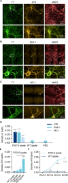
Fig 2. Triton-insoluble tau is positive for markers of tau pathology.
Representative images from hTau neurons transduced with T7-P301S LV at DIV4, treated with P301S lysates at DIV7 and fixed at DIV18. Immunofluorescence for T7 (green), MAP2 (red) and markers of tau pathology (yellow) AT8 (A), PHF-1 (B) and MC-1 (C). D) Quantification of the density of Triton-insoluble AT8/PHF-1/MC-1 spots in hTau neurons transduced with T7-P301S LV and treated with PBS, WT or P301S lysates. Data shown as mean ± SEM, n = 3 independent experiments. Two-Way ANOVA with Sidak’s multiple comparisons test. For each marker, all conditions were tested against PBS control. E) Quantification of the density of Triton-insoluble AT8 spots in cells transduced with T7-P301S LV and treated with WT lysate at 166 pg/cell, or P301S lysate at the indicated concentrations. Data shown as mean ± SEM, n = 3 independent experiments. One-Way ANOVA with Dunnett’s multiple comparisons test. All conditions compared with WT lysate. F) Quantification of the density of Triton-insoluble AT8 spots in cells transduced with T7-P301S LV and treated with WT or P301S lysate at 166 pg/cell. Data shown as mean ± SEM, n = 3–4 independent experiments. Two-Way ANOVA with Sidak’s multiple comparisons test, comparing WT vs P301S lysate for each DIV.
Fig 3
Fig 3. P301S lysate seeding potency correlates with formation of tau inclusions in seeded neurons.
A) Levels of accumulated tau, as measured by Tau aggregation HTRF assay, in brain lysates from WT, KO and various P301S animals (9 P301S lysates identified as L1-L9). For ease of comparison, raw values were normalised to the value of the P301S lysate with the lowest signal (P301S L1). Data shown as mean ± SEM, n = 2 technical replicates. B) Seeding potency measured by luminescence-based Tau Biosensor Assay using the same lysates as in (A). Raw values were normalised to the value of the P301S lysate L1. Data shown as mean ± SEM, n = 3–4 independent experiments. One-Way ANOVA with Dunnett’s multiple comparisons test. All conditions compared with WT lysate. C) Density of Triton-insoluble AT8 spots in hTau primary neurons transduced with T7-P301S LV and treated with the lysates form (A). Data shown as mean ± SEM, n = 3–4 independent experiments. One-Way ANOVA with Dunnett’s multiple comparisons test. All conditions compared with WT lysate. D-E) Linear regressions between the parameters Density of AT8 spots and Tau aggregation HTRF (D) or results of the Biosensor Assay (E). Dotted lines represent 95% confidence interval. F-G) Density of Triton-insoluble AT8 spots and representative images from hTau primary neurons transduced with T7-P301S LV and treated with a selection of lysates from (A). Cells were fixed at the time-points indicated in the graph. Data shown as mean ± SEM, n = 3 independent experiments. Two-Way ANOVA with Sidak’s multiple comparisons test, comparing WT vs P301S lysates for each DIV.
Fig 4
Fig 4. Second generation seeding.
A) Diagram of experimental design. 1st generation of hTau primary neurons were transduced at DIV4 with T7-P301S LV or Control LV at MOI 5 and incubated at DIV7 with 166.6 pg/cell of WT or P301S lysates, up to DIV18. 2nd generation of hTau primary neurons were transduced at DIV4 with T7-P301S LV at MOI 5 and incubated at DIV7 with lysates of 1st generation seeded hTau primary neurons at MOS 166.6 pg/cell, up to DIV18. B) Biosensor Assay measuring original WT and P301S lysates (brain lysates) and lysates from the 1st generation of hTau primary neurons (1st generation cells). Data shown as mean ± SEM. Brain lysates: n = 4 technical replicates. 1st generation cell lysates: n = 3 independent cultures. One-Way ANOVA with Dunnett’s multiple comparisons test. All conditions compared with WT lysate. C-D) Density of AT8 spots and representative images of 1st and 2nd generation seeded hTau primary neurons (1st generation cells and 2nd generation cells, respectively). Data shown as mean ± SEM, n = 3 independent cultures. One-Way ANOVA with Dunnett’s multiple comparisons test. All conditions compared with T7-P301S LV + WT lysate, within each generation of cells.
Fig 5
Fig 5. Anle138b prevents tau accumulation in seeded primary neurons.
A) Density of Triton-insoluble AT8 spots and representative images (B) from hTau primary neurons transduced with T7-P301S LV at DIV4 and treated with P301S lysates at DIV7, in the presence of 1 μM and 3 μM of the anti-aggregation compound Anle138b from DIV6. DMSO was used as vehicle. Data shown as mean ± SEM, n = 5 independent experiments. One-Way ANOVA with Dunnett’s multiple comparisons test. All conditions compared with DMSO control.
Fig 6
Fig 6. Human tau knock-down by siRNA decreases tau inclusions in seeded hTau primary neurons.
A) Diagram of experimental design. hTau primary neurons transduced with T7-P301S LV at DIV4 were treated with WT or P301S lysates at DIV7 and increasing concentrations of human hTau siRNA, from either DIV7 or DIV11. Non-targeting pool (NTP) siRNA at 0.5 μM was used as a control. Cells were lysed for HTRF assay or fixed for immunofluorescence at DIV18. Total tau HTRF of seeded primary neurons treated with siRNA at DIV7 (B) or DIV11 (C). Accumulated tau as measured by Tau aggregation HTRF (D-E) or Triton-insoluble AT8 spots as quantified by IF (F-G). Grey bars: treatment with T7-P301S and WT or P301S lysates, without siRNA. Blue bars: treatment with T7-P301S LV, P301S lysate and siRNA as indicated in the graph. Data shown as mean ± SEM, n = 3 independent experiments. One-Way ANOVA with Dunnett’s multiple comparisons test, all conditions compared with NTP treatment. H) Representative images from hTau neurons treated as in (F-G) with 0.5 μM of NTP or hTau siRNA.
Fig 7
Fig 7. Implementation of the tau seeding model for drug screening.
A) Diagram highlighting the key steps in the culture and treatment of primary neurons for use with the tau seeding model. Cortical neurons are cultured from hTau embryos, transduced with T7-P301S LV at DIV4 and treated with P301S lysates at DIV7. WT lysates are used as a negative control. Tau inclusions are detectable from DIV14 onwards. Treatments aimed at reducing the level of accumulated tau can be started from DIV7. The specific time-points of intervention and analysis may be defined by the end user according to experimental needs. B) Strategy for obtaining large quantities of comparable seeding material to reduce seeding variability.

References

    1. Guo T, Noble W, Hanger DP. Roles of tau protein in health and disease. Acta Neuropathol (Berl). 2017;133: 665–704. doi: 10.1007/s00401-017-1707-9 - DOI - PMC - PubMed
    1. Arendt T, Stieler JT, Holzer M. Tau and tauopathies. Brain Res Bull. 2016;126: 238–292. doi: 10.1016/j.brainresbull.2016.08.018 - DOI - PubMed
    1. Colin M, Dujardin S, Schraen-Maschke S, Meno-Tetang G, Duyckaerts C, Courade J-P, et al.. From the prion-like propagation hypothesis to therapeutic strategies of anti-tau immunotherapy. Acta Neuropathol (Berl). 2020;139: 3–25. doi: 10.1007/s00401-019-02087-9 - DOI - PMC - PubMed
    1. Lee VM-Y, Goedert M, Trojanowski JQ. Neurodegenerative Tauopathies. Annu Rev Neurosci. 2001;24: 1121–1159. doi: 10.1146/annurev.neuro.24.1.1121 - DOI - PubMed
    1. Congdon EE, Sigurdsson EM. Tau-targeting therapies for Alzheimer disease. Nat Rev Neurol. 2018;14: 399–415. doi: 10.1038/s41582-018-0013-z - DOI - PMC - PubMed

Publication types